
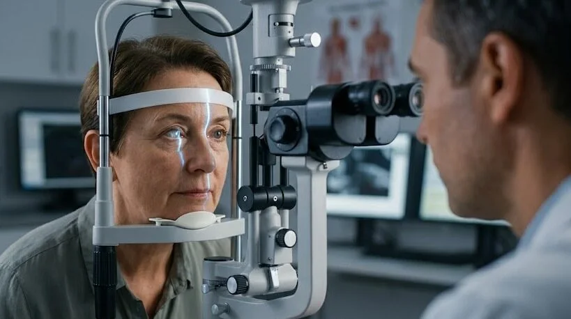
When a serious eye infection does not respond to drops or tablets, doctors may need to replace the damaged cornea. This procedure is called TPK eye surgery, or therapeutic penetrating keratoplasty. Unlike a standard corneal transplant intended primarily to improve sight, its immediate purpose is to control the infection and preserve the eye.
This guide covers who may need the procedure, how it is performed, its possible complications and what recovery may involve, helping you prepare for a discussion with your surgeon.
Key Takeaways
- The procedure replaces the infected cornea with donor tissue that extends through the full corneal thickness.
- The primary aims are to eliminate the infection and preserve the eye, rather than promise normal or sharp vision.
- Doctors generally consider it when infection does not respond to medical therapy or the cornea is at risk of perforating.
- Operating within about a week of treatment failure is linked to better graft survival.
- Glaucoma, graft rejection, reinfection and cataract are the most common complications.
- Recovery can require months of prescribed eye drops and frequent monitoring, and some patients later need another operation to improve vision.
What Is TPK Eye Surgery?
TPK is short for therapeutic penetrating keratoplasty. During the procedure, the surgeon removes the diseased portion through the full thickness of the cornea—the transparent dome at the front of the eye—and sutures donor corneal tissue into its place.
The term “therapeutic” describes the main purpose. An optical transplant is performed when the eye is stable and the goal is improved vision. With TPK, the eye is infected or inflamed, so the priorities are different: control the infection, close and protect the eye, and address vision later.
Published reviews indicate that approximately 3% to 38% of severe microbial keratitis cases eventually require transplantation. The broad range is influenced by geographic differences, the infecting organism and how quickly specialist treatment is obtained.
When Do Doctors Recommend Therapeutic Keratoplasty?
Surgery is generally not the initial treatment. It is considered when situations such as these arise:
- The cornea has perforated, or it has thinned so much that rupture is imminent.
- The ulcer keeps growing despite intensive treatment. For bacterial ulcers, no improvement after 48 to 72 hours of fortified antibiotics is a common trigger to start planning.
- Stubborn organisms such as Acanthamoeba, Pythium, microsporidia or Nocardia are involved.
- Infection is creeping toward the white of the eye or into the eye itself, raising the risk of endophthalmitis.
Fungal ulcers are more likely than bacterial ulcers to require transplantation. Studies report rates ranging from 15% to 55%, with larger and deeper ulcers, particularly those with pus behind the cornea, carrying the greatest likelihood.
Other Corneal Transplant Options and When They Fit
A full-thickness graft is not necessary for every infected cornea. When the infection has not reached the deepest layers, deep anterior lamellar keratoplasty (DALK) may be an option. This technique preserves the patient’s own inner corneal lining and can reduce rejection risk.
One comparison of advanced cases found clear grafts at one year in about 90% of DALK eyes versus 78% of full-thickness grafts. The catch is selection: fewer than one in five therapeutic cases qualify, and perforation or deep infection rules DALK out.
Less extensive approaches are also available. A conjunctival flap may help support a healing ulcer, while tissue adhesive combined with a bandage lens can close perforations smaller than about 2 mm. These measures often provide temporary support rather than eliminate the need for surgery.
When fresh donor tissue is unavailable, glycerol-preserved cornea can provide structural support. It has no living inner lining, so it protects the eye but rarely delivers good vision on its own.
Getting Ready for TPK Eye Surgery
Although TPK often has to be arranged quickly, preparation remains important. The medical team records the ulcer’s size and appearance, and an ultrasound may be performed when the infection prevents a clear view inside the eye.
Whenever possible, corneal scrapings are collected for staining and culture before surgery because identifying the organism helps determine subsequent treatment. If perforation has already occurred, the excised corneal button can instead be sent for testing.
Surgeons also look at the bigger picture. Diabetes, earlier steroid use and poor access to follow-up all affect the odds, and one review linked diabetes to roughly 2.8 times higher odds of anatomical failure.
Counselling is also an important part of preparation. Patients should understand that preserving the eye is the first priority and that additional surgery may be necessary later.
How the TPK Procedure Is Performed
General anaesthesia is most commonly used. Once the cornea is opened, the eye is temporarily an open globe, making coughing or sudden movement hazardous. Local anaesthetic blocks may be considered for cooperative adults with small peripheral ulcers, whereas children require general anaesthesia.
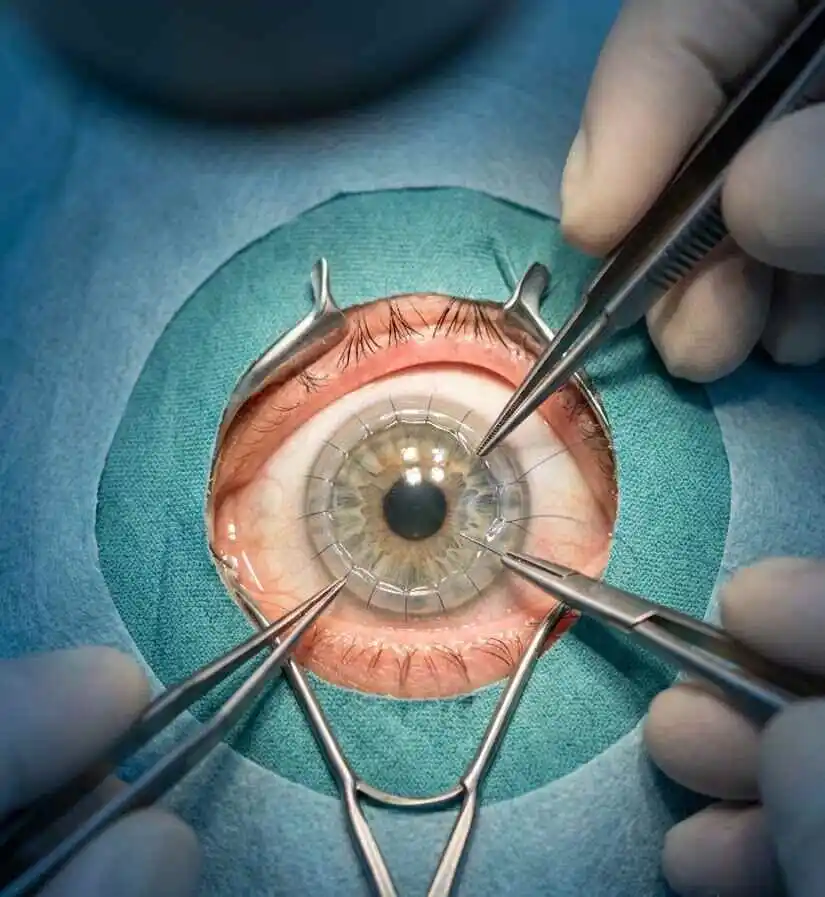
The surgeon chooses an opening large enough to remove all infected tissue together with a narrow rim of healthy cornea. The diseased corneal button is removed and the inside of the eye is irrigated. Antimicrobial medication is often administered inside the eye at this point.
Any remaining infected tissue is trimmed from the edges. If the lens is clouded or contaminated, it may be removed in the same sitting.
The donor cornea is prepared slightly larger than the opening, by about 0.5 to 1 mm, and attached with approximately sixteen fine interrupted nylon sutures. Because the sutures are separate, individual stitches can later be removed if infection returns around the graft edge.
With larger grafts, a small hole in the iris is often made to prevent a pressure-raising blockage. That step is a routine part of the TPK procedure, not a sign that something went wrong.
Risks and Complications of TPK Eye Surgery
Surgery in an inflamed eye carries greater risk than a planned elective transplant. Reported complications include:
- Recurrent infection: reported anywhere from 0% to 30%, and higher with fungal and unusual organisms.
- Secondary glaucoma: seen in roughly 20% to 43% of eyes in one review.
- Graft rejection: about 22% within the first year in one series.
- Cataract: affects around two thirds of eyes that still have their natural lens.
- Slow surface healing or graft melt, especially when preserved tissue is used.
- Endophthalmitis: uncommon, with one series reporting about 1.7%.
Larger ulcers (over 6 mm), spread to the corneal edge and delayed surgery raise the odds of reinfection. Pythium keratitis is the toughest group, with high recurrence and early graft failure in several series.
Recovery After TPK Eye Surgery
Recovery can be prolonged and intensive. Antimicrobial drops are usually given frequently at first and then reduced over the following weeks. Certain infections, including Acanthamoeba, may require treatment for several months.
Steroid drops are timed with care. Bacterial cases may start them within days, while fungal, Acanthamoeba and Pythium cases often wait until infection control is clear. Your surgeon will also check eye pressure often, because glaucoma can creep in early.
Follow-up visits are close together, often daily during week one, weekly through the first month and then monthly. Sutures commonly remain for six to twelve months because a previously infected eye heals more slowly.
After the eye has become stable, often within three to six months, visual rehabilitation may start with a rigid contact lens or adjustments to the sutures. If another optical graft is needed, it is usually postponed for at least six to twelve months.
Practical habits make a real difference: use every drop exactly as scheduled, protect the eye from knocks and never skip a review.
Success Rates and Vision Outcomes After TPK Eye Surgery
A large review pooled results from ten studies. Around 89% of eyes were preserved and about 91% had their infection cured. Only about 45% of grafts were still clear at last follow-up, and roughly 20% of eyes reached walking vision of 6/60 or better.
A single-centre study of 80 eyes tells a similar story. Infection was eradicated in 90% of fungal and 95% of bacterial cases, yet only 6% to 8% ended with a clear graft and vision better than 6/60.
The difference between preserving the eye and restoring useful sight is an important reality of this procedure. Even limited vision, such as detecting hand movements or counting fingers, may improve independence and can help avoid a painful, blind or surgically removed eye.
Why Timing Matters So Much
In the 80-eye study, graft survival at six months was 95% when surgery happened within seven days of presentation and 82.5% when it happened later. The study was retrospective, so the result shows an association, not proof.
The practical lesson remains the same: when an ulcer is clearly failing to respond, prompt referral to a corneal surgeon who can assess the need for TPK eye surgery is preferable to spending another week simply increasing drops.
Warning Signs After Surgery
Call your surgeon the same day if you notice any of the following:
- A sudden drop in vision.
- Worsening pain, redness or light sensitivity.
- New discharge, a white spot or visible swelling of the graft.
- Fluid leaking from the eye or a sense that the eye feels soft.
These symptoms may indicate reinfection, graft rejection or leakage from the wound, all of which are more manageable when identified early.
Frequently Asked Questions
What does TPK eye surgery stand for?
TPK means therapeutic penetrating keratoplasty, a full-thickness corneal transplant done mainly to treat serious infection.
Is TPK eye surgery painful?
The operation itself is done under anaesthesia, usually general. Afterwards the eye is commonly sore, watery and light-sensitive. Pain that suddenly worsens should be reported promptly.
How long does recovery take after TPK eye surgery?
Surface healing takes weeks, drops continue for months and stitches often remain for six to twelve months. Final vision may take a year or more, particularly if a second graft is needed.
What is the success rate of a therapeutic corneal transplant?
Roughly nine in ten eyes are saved and the infection cleared. Far fewer grafts stay clear long term, so success is measured by saving the eye first and vision second.
Can I see clearly again after a cornea transplant for infection?
Some people do, especially after a later optical transplant, but many are left with limited vision. Your surgeon can estimate the likely outcome from the infection type and the eye’s condition.
Bottom Line
TPK eye surgery is generally a last-resort procedure that preserves the eye in most cases, although restoring clear vision is far less certain. Prompt referral, careful postoperative treatment and realistic expectations are especially important. If a corneal ulcer is failing to improve, seek urgent evaluation by an ophthalmologist. This guide is educational and cannot substitute for individual medical advice.