

Keratoconus eye surgery is not one single operation. It is a group of procedures, ranging from a short cross-linking treatment that steadies the cornea to a full corneal transplant, and the right choice depends on how far the condition has advanced.
Many people with keratoconus never need an operation at all, because glasses or specialist contact lenses carry them for years. This guide explains when keratoconus surgery becomes necessary, what each option does, how recovery works and where the risks lie.
Key Takeaways
- Keratoconus eye surgery covers several different procedures, not one operation.
- Cross-linking aims to stop the cornea from getting worse, not to sharpen vision.
- Ring segments and CAIRS reshape the cornea to improve sight.
- A corneal transplant is usually kept for advanced disease.
- Avoiding eye rubbing protects the results of any treatment.
- Regular check-ups matter because keratoconus can keep changing.
What Is Keratoconus?
Keratoconus is a condition in which the clear front surface of the eye, the cornea, gradually thins and bulges into a cone-like shape. That uneven surface scatters light, which is why vision becomes blurred, distorted or doubled.
It usually starts in the teens or early adult years. Reported rates vary widely across the world, but one meta-analysis covering 15 countries put the global figure at about 1.4 per 1,000 people, with higher rates in younger groups.
Nobody knows the exact cause. Genetics, allergy, repeated eye rubbing and ultraviolet exposure all seem to play a part. A large observational study in the United States followed 1,209 people with keratoconus for three years to learn how the disease behaves and what drives its progression.
Common symptoms
- Blurred or distorted vision that slowly gets worse.
- Glasses or contact lens prescriptions that keep changing.
- Ghosting or multiple images, especially in one eye.
- Sensitivity to light and glare.
- Eye strain after reading or screen work.
How it is diagnosed
Your eye doctor maps the shape of the cornea with topography or tomography, measures its thickness with pachymetry and examines it under a slit lamp. Because the disease changes over time, repeat scans matter.
Doctors often judge progression by small shifts, for example when the steepest curvature of the cornea rises by about one diopter within a year.
Non-Surgical Care to Try First
Keratoconus surgery is rarely the first step. Early on, the focus is on protecting the cornea and correcting vision as well as possible.
- Stop eye rubbing and keep allergies under control, since both are linked to progression.
- Glasses or soft toric lenses for mild, early disease.
- Rigid gas permeable lenses, which create a smooth surface over the uneven cornea.
- Hybrid lenses with a firm center and a soft skirt for extra comfort.
- Scleral or mini-scleral lenses that vault over the cornea and rest on the white of the eye.
- Piggyback or specialty lens systems when other lenses are not tolerated.
When Is Keratoconus Eye Surgery Needed?
Keratoconus surgery usually enters the conversation in two situations. The first is when the cornea is clearly getting worse. The second is when vision can no longer be corrected well with glasses or lenses, or when lenses simply become unbearable to wear.
There is no single rulebook. Surgeons weigh your age, allergies, contact lens tolerance, how severe the disease is, where the cone sits, how thick the cornea is and whether there is scarring. The surgeon’s own experience also shapes the plan.
Catching the problem early matters. Keratoconus treatment that stabilizes the cornea tends to work better before the disease is advanced, and it can reduce the chance of needing a transplant later.
Main Types of Keratoconus Eye Surgery
Here is a quick overview before we go through each option in detail.
| Option | Main aim | Usually considered when |
| Corneal cross-linking | Strengthen the cornea and stop progression | Keratoconus is getting worse, ideally early |
| Intracorneal ring segments | Reshape the cornea to improve vision | Lenses are not tolerated and the cornea is clear |
| CAIRS | Reshape the cornea with donor tissue | Similar to rings, often when plastic rings are a concern |
| Combined procedures | Stabilize and smooth the cornea together | Moderate disease with irregular astigmatism |
| Corneal transplant | Replace diseased corneal tissue | Advanced disease or significant scarring |
Corneal cross-linking
Corneal cross-linking (CXL) is the keratoconus treatment most often used to stop the cornea from worsening. Riboflavin (vitamin B2) drops are applied to the cornea, which is then exposed to ultraviolet A light. The reaction forms new bonds between collagen fibers and stiffens the tissue.
The traditional method removes the surface layer first, soaks the cornea in riboflavin and then applies light. Faster versions shorten the treatment, and epithelium-on versions leave the surface layer in place to reduce discomfort and infection risk. The evidence on how well the epithelium-on versions work is mixed, so ask your surgeon which one they recommend and why.
CXL works best in early disease. A trial in young people aged 10 to 16 found that an accelerated protocol halted progression better than standard care, and Cochrane reviewers have assessed randomized trials of cross-linking against no or sham treatment, although the number of trials available was small.
Keep your expectations realistic. The goal is stability, not clearer vision, although some people notice a modest improvement as the cornea flattens.
Intracorneal ring segments
Ring segments such as Intacs are tiny curved implants, usually made of a clear plastic, placed in a channel inside the cornea. The channel can be cut by hand or with a femtosecond laser. The rings flatten the center of the cornea and make its shape more regular.
They are most useful when lenses are not tolerated, vision cannot be corrected well, and the cornea is clear and thick enough to hold the implants. They are less suitable for very advanced disease, corneal scarring, severe allergy, ongoing eye rubbing or pregnancy.
The rings can be removed, which is a real advantage. Their downsides include segments working loose or pushing through the surface over time.
CAIRS (corneal allogenic intrastromal ring segments)
CAIRS is a newer idea. Instead of plastic, the surgeon places ring-shaped segments cut from donor corneal tissue inside the cornea. Because the tissue is human, it integrates well, and it avoids the protrusion problem that has led some plastic rings to be removed.
Optometrists who work with the procedure describe better quality of vision, and in some patients a more comfortable fit with ordinary contact lenses. A small study of 24 eyes combining CAIRS with cross-linking reported improved vision and corneal shape after 18 months, with no segments displaced.
People who have not yet had cross-linking are often advised to have it a few months after CAIRS. Since the technique is still fairly new, long-term data are limited.
Combined procedures
Some surgeons combine cross-linking with another step to smooth the cornea, such as topography-guided laser treatment or ring segments. This approach is often called CXL plus.
It is worth knowing that standard refractive eye surgery such as LASIK is traditionally avoided in keratoconus, because it can weaken the cornea further. Combined treatments use laser in a carefully planned, limited way alongside cross-linking.
Corneal transplant (keratoplasty)
Up to one in five people with keratoconus may eventually need a transplant. As surgery for keratoconus goes, it is usually the last resort, used when the cornea is too thin, too scarred or too irregular for other options.
Two main types exist. Deep anterior lamellar keratoplasty (DALK) replaces the front and middle layers and keeps your own inner lining, which lowers the risk of rejection. Penetrating keratoplasty (PK) replaces the full thickness and is chosen when scarring runs deep or the cornea is extremely thin.
Both have a long track record. In an Australian registry, about 89% of penetrating grafts for keratoconus were still working at 10 years, falling to about 49% at 20 years. Younger patients may therefore need more than one graft in their lifetime.
DALK is technically harder and depends heavily on the surgeon’s experience, but it usually needs less long-term steroid use, which lowers the risk of raised eye pressure.
What is coming next
Researchers are exploring new keratoconus treatment ideas, including Bowman’s layer transplants, donor-tissue lenticules, stem cell therapy and gene-based approaches, and artificial intelligence is being studied for earlier detection. Most of this is still experimental, so treat it as hope rather than an option today.
Which Surgery Fits Which Stage?
Every eye is different, but these general patterns show how the options usually line up with the severity of the disease. They are guides, not rules.
- Early or mild: regular monitoring with glasses or soft lenses, and cross-linking if scans show the cornea is changing.
- Moderate: cross-linking, often with ring segments, CAIRS or a combined procedure, alongside specialty lenses if needed.
- Advanced or scarred: a corneal transplant, usually after other options have been considered or have not been enough.
Surgery is generally on the table when the cornea has scarred, has become extremely thin, or when even the strongest lenses cannot give usable vision. It is also considered when you simply cannot tolerate any type of contact lens.
Keratoconus in Children and Teens
Keratoconus often begins in the teenage years, and it is reported to be more common in younger groups than once believed. In young people it can also move faster, which is why early detection matters.
It is worth asking for a corneal scan if a child has a fast-changing prescription, rubs their eyes a lot, has allergies or has a close relative with keratoconus. A trial in 60 young people aged 10 to 16 showed that accelerated cross-linking can halt progression, so treatment does not have to wait until adulthood.
Ring segments, by contrast, are generally placed in adults aged 18 and over.
Before Keratoconus Eye Surgery: What to Expect
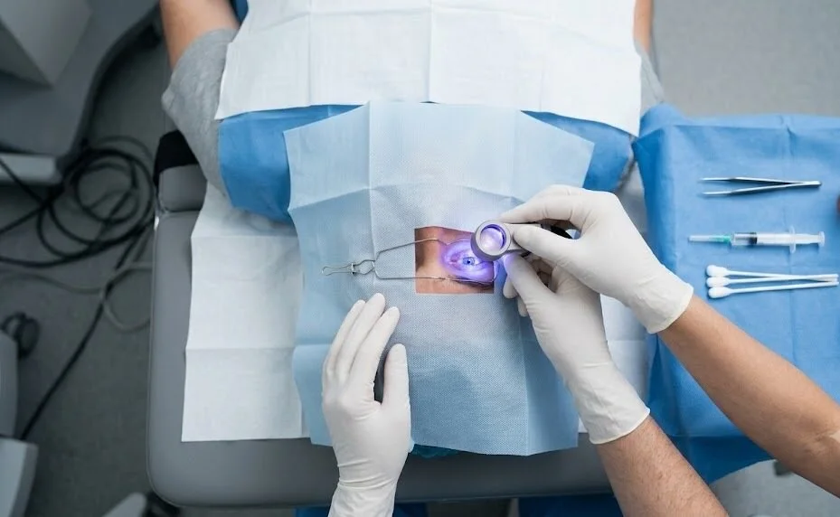
Your surgeon will start with a detailed examination and fresh corneal scans to confirm the stage of the disease and the best procedure.
- A review of your medical history, allergies and medicines.
- Corneal topography or tomography, plus thickness measurements.
- A conversation about your work, lifestyle and what you hope to see better.
- Advice on lenses, since you may be asked to stop wearing them before scans.
- Treatment for allergies or dry eye so the surface is in good shape.
Most of these procedures use numbing drops or local anesthesia, so you stay comfortable while awake. Transplants take longer and may involve different anesthesia, which your surgeon will explain.
Recovery After Keratoconus Eye Surgery
Recovery from surgery for keratoconus depends on the procedure. Cross-linking and ring procedures generally settle over days to weeks, while a transplant can take months, and vision may keep fluctuating for a while.
After epithelium-off cross-linking, the eye often feels gritty, watery and light-sensitive for a few days while the surface heals. The shape of the cornea also continues to change gradually for up to a year.
These habits help in almost every case:
- Use the prescribed antibiotic and anti-inflammatory drops exactly as directed.
- Wear sunglasses or protective eyewear outdoors.
- Do not rub or press on the eye, ever.
- Avoid swimming, dust and heavy work until your surgeon says it is safe.
- Go to every follow-up visit, because stitches or lenses may need adjusting.
Contact your surgeon right away if you have severe pain, a sudden drop in vision, increasing redness, discharge or worsening light sensitivity.
Risks and Complications of Keratoconus Eye Surgery
Every type of surgery for keratoconus carries some risk, and the risks differ from one option to the next.
- Cross-linking: infection of the cornea, reactivation of herpes eye disease, corneal haze or scarring, and, rarely, damage to the inner lining or a sudden swelling called hydrops.
- Ring segments: segments moving, poking through the surface or breaking, infection, and halos or glare at night.
- Corneal transplant: graft rejection, graft failure, high astigmatism, raised eye pressure linked to long-term steroid drops, and return of the cone shape at the graft edge years later.
Most problems are manageable when caught early. That is the main reason follow-up visits are not optional.
One complication deserves special mention: acute hydrops. It happens when a tear in a deep layer of the cornea lets fluid flood in, causing sudden clouding and swelling. It is rare, and it can occur with or without surgery.
Warning signs include a sudden drop in vision, a milky or hazy look to the eye, pain, redness and strong light sensitivity. Hydrops often settles on its own over time, but it can leave a scar, so see an eye doctor urgently.
Protecting Your Results Long Term
Even successful keratoconus surgery does not close the chapter. Eye rubbing and untreated allergy are among the best-known drivers of progression, so controlling both protects your investment.
Expect regular scans, and expect to be refitted for glasses or lenses once the eye has healed. Keratoconus treatment is often a long-term process rather than a one-time fix.
What is the long-term outlook?
Keratoconus does not usually cause complete blindness. Left unchecked, though, advanced disease can seriously limit sight. With modern treatment, most people keep useful vision, especially when the condition is found and managed early.
If you also develop cataracts
Cataracts can occur in people with keratoconus, and surgery is more demanding because an irregular cornea makes lens power calculations less predictable. Careful measurements, realistic expectations and an experienced surgeon make a real difference.
Cost and Insurance Coverage
The price depends on the procedure, the clinic, the surgeon’s experience, the country and whether donor tissue is involved. A corneal transplant is a larger undertaking than a cross-linking session, so the bills are rarely comparable.
Because these treatments protect sight rather than improve appearance, they are often treated as medically necessary. Coverage still varies a great deal between insurers and countries, and some plans treat newer techniques as experimental.
Before booking, ask the clinic for a written estimate that lists tests, the procedure, any donor tissue, medicines and follow-up visits. Check with your insurer in advance, and ask whether pre-approval is needed.
Questions to Ask Your Surgeon
- Why is this option right for my stage of keratoconus?
- What result should I realistically expect, and what is the goal?
- How many of these procedures do you perform?
- What are the main risks for my eye?
- How long until I can work, drive and wear lenses again?
- Will I still need glasses or special contact lenses afterward?
Frequently Asked Questions
Does everyone with keratoconus need surgery?
No. Many people manage well for years with glasses or specialty lenses. Surgery for keratoconus is considered when the cornea is progressing or vision cannot be corrected well.
Which keratoconus eye surgery is best?
There is no single best option. Cross-linking is used to stop progression, rings or CAIRS reshape the cornea, and a transplant is reserved for advanced cases. Your surgeon chooses based on your scans and symptoms.
Does keratoconus eye surgery hurt?
The procedures themselves use numbing drops or local anesthesia, so you should feel pressure rather than pain. Some scratchiness or discomfort in the first days is common, especially after cross-linking.
How long is recovery after keratoconus eye surgery?
It varies. Cross-linking and ring procedures usually settle over days to weeks, while a corneal transplant can take months to reach stable vision.
Can keratoconus come back after surgery?
Cross-linking is designed to stop progression, but regular monitoring is still needed. After a transplant, the cone shape can sometimes return at the edge of the graft years later.
Bottom Line
Keratoconus eye surgery gives you real options, from stabilizing the cornea early to rebuilding it in advanced cases. The best results come from catching the condition early, avoiding eye rubbing and choosing a surgeon who explains why a procedure suits your eye.
If you suspect keratoconus, or your prescription keeps changing, book a corneal scan. This article is general information and does not replace an eye examination.