
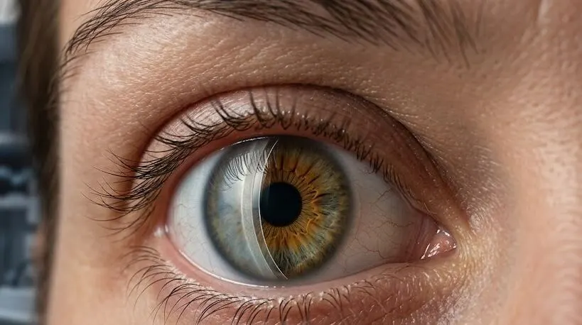
If your cornea has become cloudy and your doctor has recommended DSAEK eye surgery, you may have several concerns. Is the procedure safe? How clear will your vision become? How long will the results last? These are important questions to discuss.
DSAEK eye surgery replaces the damaged inner portion of the cornea rather than the entire cornea. It uses a small incision, with an air bubble supporting the graft, and vision generally becomes useful sooner than it does after an older full-thickness transplant.
This guide explains who may need DSAEK eye surgery, what happens during the procedure, what recovery can be like, the main risks, and how long a donor graft may remain effective.
Key Takeaways
- A DSAEK corneal transplant replaces damaged inner corneal layers with a thin disc of healthy donor tissue while leaving the front portion of your own cornea intact.
- It is commonly performed for Fuchs’ dystrophy, corneal swelling following cataract surgery, and previously failed grafts.
- The operation generally takes less than an hour, and the graft itself does not require stitches.
- Vision is usually blurred at first. It gradually becomes clearer over the following weeks and months and may continue improving for as long as a year.
- Important risks of DSAEK eye surgery include graft detachment, rejection, increased eye pressure, and gradual loss of donor endothelial cells.
- Long-term outcomes are generally favorable, but a graft does not last forever in every patient, making lifelong follow-up important.
What Is DSAEK Eye Surgery?
The cornea is the transparent front window of the eye. Its deepest layer, called the endothelium, acts like a small pump that continually removes fluid and helps keep the cornea clear and thin.
Endothelial cells cannot regenerate. When their number becomes too low, the pumping function weakens, fluid accumulates, and the cornea becomes swollen and cloudy. This can cause blurred vision, increased glare, and eventually make eye drops insufficient.
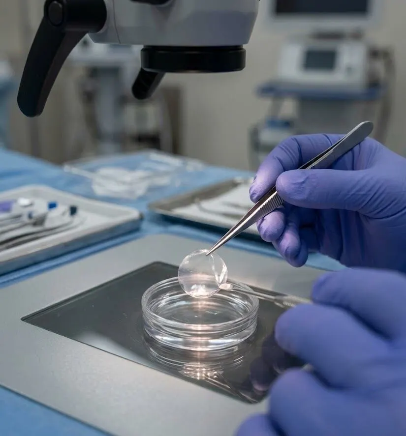
DSAEK means Descemet’s stripping automated endothelial keratoplasty. During the operation, the surgeon removes the unhealthy inner layer and replaces it with a very thin donor tissue disc containing functioning endothelial cells. A microkeratome is used to prepare the disc to the desired thickness, which is why the procedure is called automated.
This type of partial-thickness corneal transplant is part of a group of procedures known as endothelial keratoplasty. DSEK is another term you may encounter; in that technique, the donor disc is prepared by hand. The basic treatment concept is similar.
Who Needs DSAEK Eye Surgery?
The procedure is intended for people whose disease affects the endothelium while the more superficial corneal layers remain healthy. Common reasons include:
- Fuchs’ endothelial dystrophy — an inherited condition that commonly becomes noticeable after age 50. Vision is often more blurred in the morning and may improve later in the day.
- Bullous keratopathy — corneal swelling that can occur after cataract surgery or another eye procedure has injured the endothelium.
- A previously failed graft — when an earlier corneal transplant is no longer functioning properly.
- Less common causes include iridocorneal endothelial syndrome and endothelial injury caused by trauma.
Doctors generally consider transplantation when drops and other treatments no longer keep the swelling under control and it is interfering with everyday life. Delaying treatment for too long may also be unhelpful. Some research indicates that treating cataract-surgery-related swelling earlier may lead to better final vision.
A DSAEK corneal transplant cannot correct scars on the front of the cornea, keratoconus, or an irregular corneal surface. These conditions may require another treatment, which is why a detailed corneal examination is important before surgery is scheduled.
How the DSAEK Procedure Works
Most patients return home the same day. The procedure generally follows these steps:
- Anesthesia. The eye is numbed with drops or an injection around the eye, often with light sedation. General anesthesia may be used in some cases.
- Removing the damaged layer. Through a small incision, the surgeon removes the diseased endothelium along with its membrane.
- Placing the donor disc. The prepared thin donor disc is folded, inserted into the eye, and then unfolded into position.
- Air bubble. An air bubble presses the donor disc against the back surface of the cornea, allowing it to attach without stitches.
- Closing up. The small incision is closed or sealed, and an eye shield is placed over the eye.
The DSAEK procedure generally lasts about 30 to 60 minutes and may take longer when cataract surgery is performed at the same time. In some cases, the surgeon creates a very small opening in the iris beforehand to reduce the chance of pressure problems caused by the air bubble.
Recovery After DSAEK Eye Surgery: What to Expect
The first few days
For much of the first day or two, and sometimes longer, you may need to remain flat on your back so the air bubble can keep the graft against the cornea. Brief breaks for eating and using the bathroom are usually allowed. Follow your surgeon’s specific positioning instructions carefully because they help the graft remain attached.
Vision may remain hazy while the air bubble is present. The bubble gradually becomes smaller and usually disappears within several days. You will generally wear an eye shield at night and begin antibiotic and steroid eye drops.
The first few weeks
Avoid rubbing or putting pressure on the eye, and prevent soap and water from entering it. Walking is usually acceptable. Heavy lifting, strenuous exercise, swimming, and exposure to dusty environments should generally be avoided for several weeks or for the period your surgeon recommends.
Many people can return to comfortable everyday activities within roughly two to three weeks after DSAEK surgery, although their vision may still be changing.
Vision over the following months
Vision commonly becomes noticeably clearer during the first three months and may continue to improve gradually until about one year. Many patients achieve around 20/25 to 20/40 vision with glasses. The outcome varies with the duration of corneal swelling, the condition of the rest of the eye, and the thickness of the donor tissue.
DSAEK can shift the glasses prescription somewhat toward farsightedness, so it is usually better to wait for the surgeon to confirm that your prescription has stabilised before getting new glasses.
Steroid drops are generally tapered gradually over several months, and some surgeons continue a small maintenance dose for the long term to help protect the graft. Follow-up appointments are usually more frequent initially and eventually become yearly lifelong checks.
How Successful Is DSAEK Eye Surgery?
In general, results are very favorable during the early years, although outcomes vary more over a ten-year period. The reason a DSAEK corneal transplant was required has a major influence on long-term success.
One ten-year series presented at a European eye surgery meeting found that approximately 87 percent of grafts remained functional after a decade. A large German study involving eyes with Fuchs’ dystrophy reported about 73 percent survival at ten years. Other studies have found lower survival, especially in eyes affected by glaucoma or swelling after cataract surgery.
Loss of endothelial cells is an important part of the long-term picture. About one-third of donor endothelial cells may be lost during the first months, partly because the tissue is manipulated and folded during surgery. The rate of cell loss then becomes slower and more gradual.
The Cornea Donor Study reported that early endothelial cell loss after this type of endothelial keratoplasty was higher than after a full-thickness graft, but the decline became more stable sooner. This helps explain why the faster recovery and preservation of stronger corneal tissue can still make DSAEK valuable despite the initial cell loss.
Risks and Complications to Know About
Like any operation, DSAEK eye surgery has possible complications. Many of these problems can be treated successfully when they are identified promptly.
- Graft detachment. This is the most common early complication following DSAEK surgery. If the donor disc separates, the surgeon may place another air bubble, often during an office or clinic procedure. Reported rates differ considerably among surgeons and studies.
- Graft rejection. The immune system can recognize the donor tissue as foreign. Possible signs include redness, sensitivity to light, blurred vision, and aching, although early rejection may produce few noticeable symptoms. Across ten years, studies have reported rejection in roughly one in eight to one in five eyes. Prompt steroid treatment can often preserve the graft.
- Raised eye pressure. Steroid drops may increase pressure inside the eye, while the air bubble can temporarily interfere with fluid drainage soon after surgery. Eye pressure is therefore monitored during follow-up visits.
- Late graft failure. Donor endothelial cells gradually decrease over time, and some grafts eventually stop functioning. Another transplant can be considered if this happens.
- Infection and bleeding. These complications are uncommon, in part because the surgical incision is small.
- Cataract progression. In eyes that still have their natural lens, cataract formation may progress more quickly, with steroid treatment being one possible contributing factor.
DSAEK vs DMEK vs Full-Thickness Transplant
Your surgeon may discuss alternatives to a DSAEK corneal transplant. The table below summarizes the main differences.
| DSAEK | DMEK | Full-thickness (PK) | |
| Layers replaced | Endothelium, membrane and a thin layer of stroma | Endothelium and membrane only | All corneal layers |
| Stitches in the graft | None | None | Yes |
| Visual recovery | Weeks to months | Often the fastest | Slowest, sometimes over a year |
| Tissue handling | Sturdier, easier to place | Very delicate, trickier | Straightforward, but a larger wound |
| Often chosen for | Most endothelial problems and complex eyes | Fuchs’ in otherwise straightforward eyes | Scarred or misshapen corneas |
DMEK often provides somewhat sharper vision and can produce faster visual recovery, but it requires greater surgical skill and may have a higher risk of detachment. DSAEK eye surgery is still a dependable option, particularly for eyes with previous surgery or cases that are technically more challenging. The most suitable technique depends on the individual eye rather than simply choosing the newest procedure.
When to Call Your Eye Doctor Straight Away
Contact your eye doctor the same day if you develop any of the following after surgery:
- A sudden drop or clouding of vision
- Increasing redness or pain
- New sensitivity to light
- Discharge, or a feeling that the eye is getting worse rather than better
Do not wait until your next routine appointment. Problems such as rejection and graft detachment are much easier to manage when treatment begins early.
Frequently Asked Questions
Is DSAEK eye surgery painful?
The operation itself should not be painful because the eye is anesthetized. Afterward, people commonly notice a gritty or scratchy sensation, watering, and sensitivity to light for several days. Significant pain is not expected and should be reported promptly.
How long does it take to recover from DSAEK?
Most everyday activities feel normal again within about two to three weeks. Vision generally continues to improve for three to six months and may keep improving for up to a year. The final glasses prescription is usually determined after this period.
What is the success rate of DSAEK eye surgery?
Many grafts remain clear for years. Published studies report ten-year graft survival of approximately 60 to 87 percent, with the range depending on the patient population and underlying disease. Outcomes are generally strongest in Fuchs’ dystrophy.
Is DSAEK better than DMEK?
There is no single best option for every patient. DMEK can provide sharper vision and faster recovery, while DSAEK uses more robust tissue, is generally easier to position, and may detach less often. The choice is based on the condition of the eye and the surgeon’s experience.
Can a DSAEK graft be rejected, and what are the signs?
Yes. A DSAEK graft can be rejected months or even years after surgery. Warning signs include redness, sensitivity to light, blurred vision, or aching. Early rejection often responds well to steroid drops, so symptoms should be reported without delay.
Bottom Line
DSAEK eye surgery is a well-established treatment for restoring a cloudy cornea when its inner layer has stopped functioning. DSAEK surgery uses a small incision, does not require stitches in the graft, and generally allows faster visual recovery than a full-thickness transplant. However, it still carries risks, and donor endothelial cells gradually decline over time.
If your surgeon has recommended the procedure, ask why they favor that technique, what outcome is realistic for your particular eye, and how often follow-up will be needed. Taking your prescribed drops consistently and attending scheduled appointments are important for protecting the graft over the long term.