
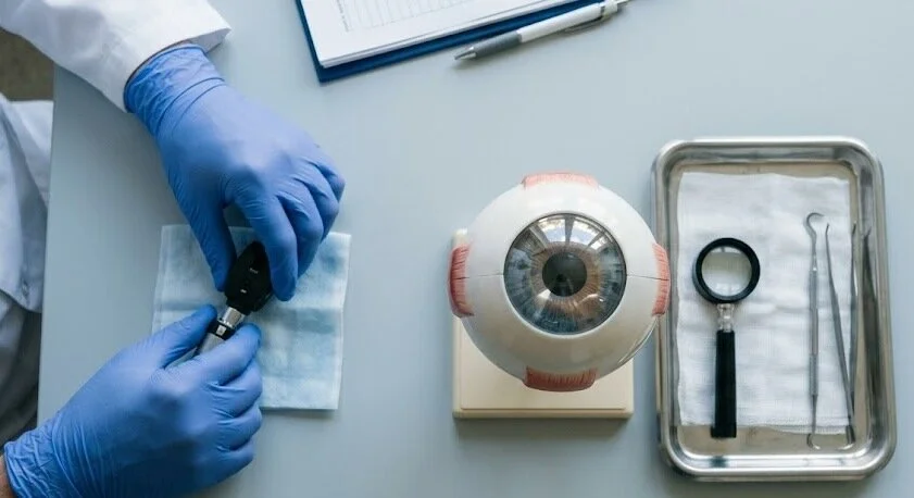
Cornea eye surgery sounds intimidating, mostly because it happens on the clear front window of your eye. Yet for many people it is the operation that finally brings back sharp vision, or ends the constant pain of a swollen, damaged surface.
This guide explains who needs the operation, how the different procedures compare, what recovery really feels like, and how often things go wrong. It is based on guidance from Mayo Clinic and Cleveland Clinic, a peer-reviewed review in the Indian Journal of Medical Research, and current clinical references.
Key Takeaways
- Most cornea eye surgery means a transplant, where damaged tissue is replaced with screened tissue from a deceased donor.
- Surgeons now try to replace only the diseased layer (DALK, DSAEK or DMEK) instead of the full thickness, which usually means fewer stitches and quicker healing.
- Cornea eye surgery typically takes under two hours and is done on one eye at a time.
- Vision can take about three months to settle after an inner-layer graft, and up to a year after a full-thickness graft.
- Rejection is the main long-term worry. Eye pain, redness, light sensitivity or hazy vision means you should call your surgeon straight away.
- Check-ups continue for life, because graft risks do not disappear after the first year.
What Is a Cornea Transplant, Really?
Your cornea is the clear, dome-shaped layer at the front of the eye. It bends incoming light and does most of the focusing before light even reaches the lens. When it turns cloudy, scarred, swollen or misshapen, the image on the retina blurs, and glasses often cannot fix that.
Surgery swaps the damaged tissue for healthy donor tissue. Sometimes the whole thickness is replaced, and sometimes only a single thin layer. Surgeons call this keratoplasty, while most patients simply say they are having a cornea transplant.
It is one of the most common transplants in the world, with roughly 185,000 operations a year, according to Cleveland Clinic. The cornea has no blood vessels, which is a big reason your immune system usually tolerates the graft.
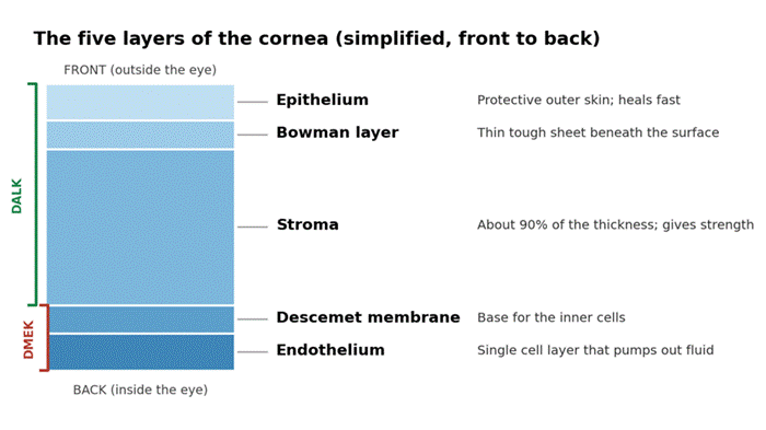
Figure 1: The cornea has five main layers. Surgeons can replace only the layers that are diseased.
Who Needs Cornea Eye Surgery?
Not every cloudy cornea needs an operation. Doctors usually try drops, special lenses and other treatments first. Corneal surgery comes into the picture when vision loss, pain or swelling cannot be controlled any other way. Common reasons include:
- Keratoconus, where the cornea thins and bulges into a cone shape.
- Fuchs dystrophy, an inherited condition in which the inner cell layer slowly stops working.
- Scars left by infections, injuries or old ulcers.
- Bullous keratopathy, a painful blister-like swelling that can follow other eye surgery.
- Corneal ulcers that do not respond to medicine.
- A previous graft that has failed.
The mix differs around the world. A ten-year review of US data found Fuchs dystrophy had become the leading reason for grafts, while in many developing regions infections and scarring still dominate.
The Main Types of Cornea Eye Surgery
Modern surgeons try to replace only the layer that is diseased. That keeps more of your own tissue and often lowers the rejection risk. In that same US review, full-thickness grafts fell from about 95% of cases in 2005 to around 42% in 2014, as partial-thickness keratoplasty took over.
| Procedure | What is replaced | Often used for | Worth knowing |
| Penetrating keratoplasty (PKP) | The full thickness of the central cornea | Deep scars, damage across all layers, failed grafts | Needs stitches; slowest visual recovery |
| Deep anterior lamellar (DALK) | Front and middle layers; your inner layer stays | Keratoconus, scars with healthy inner cells | Eye is not opened fully, so rejection risk is lower |
| DSAEK / DSEK | Inner cell layer plus a thin layer of support tissue | Fuchs dystrophy, bullous keratopathy | Held in place by an air bubble; few or no stitches |
| DMEK | Only the thinnest inner membrane and cells | Fuchs dystrophy and similar problems | Fastest visual recovery, but a graft may need re-bubbling |
| Superficial lamellar (SALK) | Only the front surface | Shallow scars | Sometimes done without stitches |
| Artificial cornea (keratoprosthesis) | The cornea is replaced by a device | Repeated graft failure or very high-risk eyes | Reserved for difficult cases |
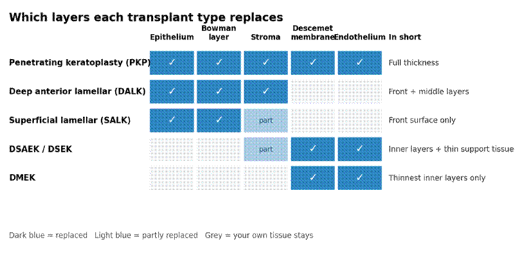
Figure 2: Which layers each type of graft replaces, and which of your own layers stay in place.
Which option suits you depends on how deep the damage runs and whether your innermost cells still work. Your surgeon will usually decide this after a close examination and imaging, before a date is set.
Preparing for Surgery and Where Donor Tissue Comes From
Before cornea eye surgery, expect a detailed eye exam, measurements to size the graft, and a review of your medicines and supplements. Other problems, such as infection or inflammation, are usually treated first because they can hurt the graft’s chances.
Fasting note: Your surgical team will tell you exactly when to stop eating and drinking before the operation, and which medicines to pause or keep taking. Instructions differ depending on the type of anesthesia, so follow the written plan you are given rather than a general rule. Arrange a driver and, if possible, someone to stay with you that evening.
Donor corneas come from people who have died. Eye banks screen every donor, and tissue is rejected if the cause of death is unknown or if the donor had certain infections or earlier eye surgery. Unlike a kidney or liver, a cornea does not have to be matched to you.
In the United States the wait is usually short. In many other countries supply is the bottleneck. Cleveland Clinic reports only one cornea is available for every 70 people who need one, and people in more than half of the world’s countries cannot get this surgery at all.
What Happens on the Day of Cornea Eye Surgery
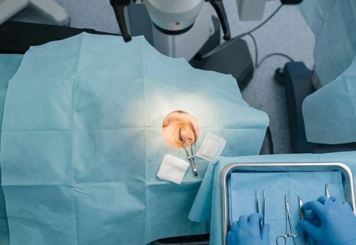
You will receive either sedation with a numbing medicine for the eye, or sometimes general anesthesia. Either way, you should not feel pain. One eye is treated at a time, and most operations take under two hours.
In a full-thickness graft, the surgeon removes a round button of diseased cornea and stitches the donor piece into the opening. In inner-layer operations, a very thin donor layer goes in through a small cut and is pressed into place with an air bubble, so far fewer stitches are needed.
Most people go home the same day with a protective shield. You will need someone else to drive you.
Recovery After Cornea Eye Surgery
Expect a red, scratchy, light-sensitive eye for the first few days. Your surgeon will usually see you within a day or two to check healing and remove the patch. After that, recovery follows a fairly predictable routine:
- Eye drops: antibiotic drops prevent infection, while steroid drops calm inflammation and help protect against rejection. Anti-rejection drops often continue for at least a year.
- Positioning: after an inner-layer graft you may need to lie face up for a few days so the air bubble keeps the tissue in place.
- Protection: wear the shield or glasses as directed, and never rub or press the eye.
- Stitches: if you have them, some may stay in for several months and are removed gradually.
- Follow-ups: visits are frequent in the first year, then yearly for life.
Vision often gets blurrier before it gets better. After DMEK or DSAEK, many people see well within about three months, while full-thickness grafts can take up to a year to settle. Glasses, contact lenses or sometimes a laser touch-up may be needed to sharpen the result after corneal surgery, especially if stitches have left astigmatism.
Returning to work can take a few days to a few weeks, depending on your job. Desk work usually comes back sooner than heavy lifting. Driving needs your surgeon’s approval, because advice and local rules vary.
Risks, Rejection and Warning Signs
A corneal transplant is generally safe, but it is not risk free. Possible problems include infection, high eye pressure (glaucoma), bleeding, stitch trouble, retinal swelling or detachment, and a graft that slips out of position. A slipped inner-layer graft can often be fixed by injecting another air bubble.
Rejection is the one most people worry about. It happens when your immune system treats the donor tissue as foreign. Mayo Clinic puts the overall rate at around 10%, though reported figures vary by technique, and Merck Manuals lists roughly 1% to 3% for DMEK.
Caught early, rejection can usually be treated with steroid drops. Call your surgeon the same day if you notice:
- New or worsening eye redness.
- Eye pain.
- Sensitivity to light.
- Hazy, cloudy or dropping vision.
Please do not wait to see whether it settles on its own. Early treatment is what protects the graft.
How Well Does It Work? Success Rates and Longevity
The results of a corneal transplant depend more on the diagnosis than on any single technique. Keratoconus grafts tend to do very well. Eyes with severe dryness, new blood vessels in the cornea or active infection carry a higher risk. For example, Merck Manuals cites about 97% graft survival ten years after DALK for keratoconus.
For full-thickness grafts in keratoconus, long-term studies summarized in the IJMR review put failure at roughly 6% to 7% around twenty years later.
Even so, a graft is not a lifetime guarantee. Rejection risk and slow loss of the inner cells continue for years, which is why yearly check-ups matter. If a graft does fail, a repeat transplant or, in difficult cases, an artificial cornea is still an option.
How to Protect Your Corneas
Not every corneal problem can be prevented, but many of the injuries and infections that end in a transplant can. A few habits go a long way:
- Wear protective eyewear: use safety glasses or goggles for sports, power tools, yard work, and any task involving chemicals or flying debris.
- Keep contact lenses clean: wash and dry your hands before handling them, replace lenses and cases on schedule, and never rinse them in tap water. Take them out before swimming or showering, and do not sleep in them unless your eye doctor has approved it.
- Treat infections early: redness, pain, discharge, light sensitivity or sudden blurriness needs an eye doctor the same day. Avoid using leftover drops or home remedies, which can make a corneal infection worse.
- Keep up with regular eye exams: conditions such as keratoconus and Fuchs dystrophy are easier to manage when they are caught early.
- Do not rub your eyes: constant rubbing is linked to damage and can worsen keratoconus.
- Look after your general health: avoid smoking, and ask your doctor about dry eye, which can stress the cornea.
Cost and Insurance Coverage
The total price varies widely by country, hospital, surgeon and the type of graft. It usually bundles the surgeon’s fee, the operating room, anesthesia, processing of the donor tissue, and follow-up visits. The donated tissue itself is not sold. Fees cover recovery, testing and preparation by the eye bank.
When the operation is medically necessary, health insurance or national health programs commonly cover it, though rules and out-of-pocket costs differ. Before you book, it helps to:
- Ask for a written estimate and check what it includes, such as drops, suture removal and any repeat air bubble.
- Ask your insurer whether prior approval is required.
- Check whether the surgeon and the facility are both in your network.
- Ask the hospital about price estimates and financial assistance.
What’s New in Corneal Surgery
- Intraoperative OCT: live cross-section imaging helps surgeons check graft position and depth while they operate.
- Femtosecond lasers: they cut donor and host tissue with matching edges, which can reduce astigmatism.
- Descemet stripping only: for selected people with Fuchs dystrophy limited to the center, the diseased inner layer is peeled away without a donor graft. It is not suitable for everyone.
- Bioengineered corneas: lab-made implants are being tested, but the studies so far are small and early.
Frequently Asked Questions
Is cornea eye surgery painful?
Not during the operation, since you are numbed or sedated. Afterward the eye feels gritty, sore and sensitive to light for a few days, and your surgeon may suggest an over-the-counter pain reliever. Pain that is severe or getting worse should be reported quickly, because it can signal infection or rejection.
How long does it take to recover from cornea eye surgery?
The first few days are the roughest. Most people return to light work within days to weeks, though your surgeon sets the timeline. Vision typically settles in about three months after an inner-layer graft and up to a year after a full-thickness one.
What is the success rate of a corneal transplant?
It is high, and it varies by condition. Mayo Clinic calls most operations successful, with rejection in roughly 1 in 10 cases. Merck Manuals cites around 97% ten-year survival for DALK in keratoconus. Higher-risk eyes do worse, so ask your surgeon for figures that match your diagnosis.
How long does a cornea transplant last?
Many grafts last decades, especially in keratoconus. Long-term data show about 6% to 7% failing by twenty years in that group. In other conditions, grafts may fail sooner, and a second transplant is possible.
Can you have cornea surgery without a donor cornea?
Sometimes. An artificial cornea (keratoprosthesis) is used when donor tissue is unsuitable or earlier grafts keep failing. A few people with early Fuchs dystrophy may qualify for Descemet stripping only, which needs no graft. Lab-grown corneas remain experimental.
Bottom Line
Cornea eye surgery has changed a great deal in the past two decades. Replacing only the damaged layer means smaller operations, fewer stitches and faster visual recovery for many patients, and success rates are among the best in transplant medicine.
If you are considering cornea eye surgery, ask your surgeon which layer is affected, which technique they recommend and why, and what your personal rejection risk looks like. Keep every follow-up, use your drops exactly as prescribed, and act fast on warning signs.
This article is general information and does not replace advice from your eye doctor. And if you are not already registered as an eye donor, consider doing so, since donated tissue is what makes this surgery possible.