
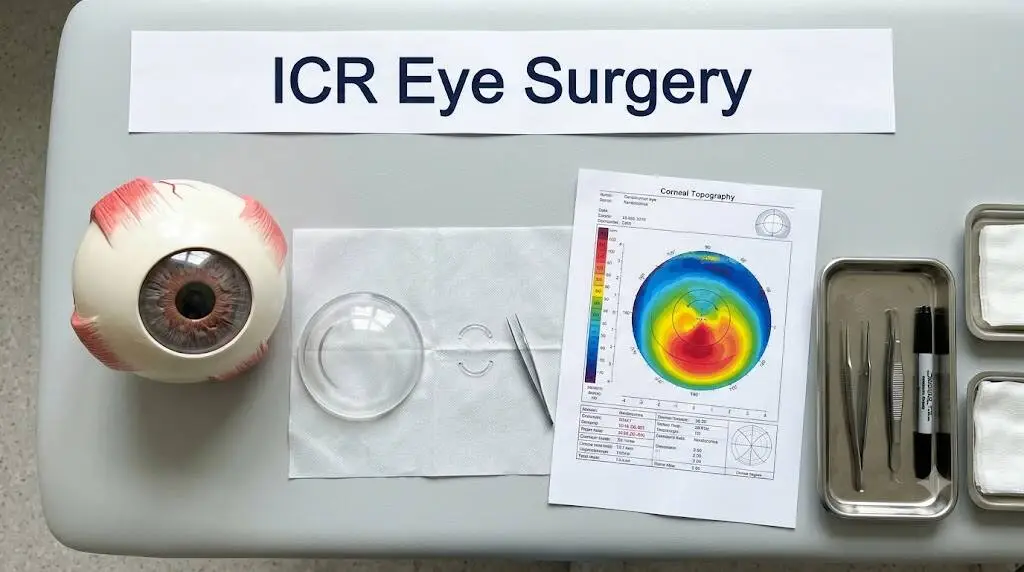
ICR eye surgery is a procedure in which a surgeon places tiny, curved plastic segments inside the cornea to smooth out its shape. It is used mainly for keratoconus, a condition where the clear front of the eye slowly thins and bulges into a cone. If glasses and contact lenses no longer give you sharp, comfortable vision, this surgery can sit in a useful middle ground between lenses and a full transplant. This guide covers how corneal ring implants work, who qualifies, what recovery feels like and what the evidence shows.
Key Takeaways
- The implants reshape the cornea. They do not remove tissue or replace it.
- People with clearly reduced vision benefit most. Those who already see well are more likely to lose vision.
- Keratoconus should be stable before surgery, because the rings alone may not stop a progressing cone.
- Serious complications are uncommon, especially when a femtosecond laser creates the tunnel.
- You may still need glasses or contacts afterward, and the segments can be removed if necessary.
What Is ICR Eye Surgery?
ICR stands for intracorneal ring. During the operation, small arc-shaped or ring-shaped implants are placed inside the middle layer of the cornea, known as the stroma. The central zone you look through is left clear.
Doctors also call these devices intracorneal ring segments, or ICRS. You may hear brand names such as Intacs, Keraring and Ferrara rings.
The operation is done under local anesthesia on an outpatient basis. Its goal is to strengthen the cornea structurally and change its curvature. It is used most often for keratoconus, but surgeons also consider it for irregular astigmatism and corneal ectasia.
How Intracorneal Rings Reshape the Cornea
In keratoconus the cornea thins and steepens, and that distorts the way light reaches the retina. The implants act like an internal scaffold. They add support, flatten the steepest area and make the surface more regular.
Early laboratory work revealed a simple rule. A thicker implant changes the cornea more, while a wider diameter changes it less. Surgeons apply this logic when they pick the number, thickness and arc length of the segments, usually guided by the manufacturer’s nomogram.
The effect can be measurable. In a five-year study of stable keratoconus, the average corneal curvature dropped by about 3.24 diopters within six months.
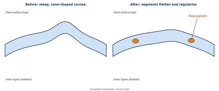
Figure 1. How ring segments change the shape of a cone-shaped cornea (illustration).
A Quick Look at How the Technique Evolved
The idea is older than most people expect. Experiments in rabbits during the 1960s showed that implants inside the cornea could change its focusing power. The concept of an intrastromal ring was formally proposed in 1978, and the first designs were full 360-degree rings aimed at nearsightedness.
Human testing began in 1991, but laser vision correction soon overshadowed the technique. It found new life in 2000, when researchers reported the first results in keratoconus. In the United States, Intacs gained FDA approval in 1999 for mild nearsightedness and a Humanitarian Device Exemption in 2004 for keratoconus.
Types of Corneal Ring Segments
Not all segments are alike. Most are made of PMMA, a clear and well-tolerated plastic, but their shapes and sizes differ. A recent review compared the main types of intrastromal corneal ring segments for outcomes and complications.
| Ring type | Design notes | Worth knowing |
| Intacs | Crescent-shaped PMMA segments with a 7 mm optical zone; Intacs SK is the 6 mm version | Best known in the U.S. |
| KeraRing | PMMA with a triangular cross-section, made for keratoconus | Customizable, though segment migration was reported more often |
| Ferrara Ring | PMMA with a triangular cross-section, first used in keratoconus in 2003 | Strong flattening in central cones, with a moderate risk of visual disturbances |
| MyoRing | The only full 360-degree design; 5 to 8 mm optical diameter | Reduced aberrations in advanced cases but a higher reoperation rate |
| CAIRS | Segments cut from donor corneal tissue | A newer, biocompatible alternative |
No single design suits everyone. The choice depends on the shape of your cone, how advanced it is and your surgeon’s experience, so ask why a particular type was selected for you.
Who Is a Good Candidate for ICR Eye Surgery?
Surgeons usually consider the procedure for keratoconus, pellucid marginal degeneration, keratoglobus and ectasia that develops after LASIK. The common thread is a distorted cornea that glasses and contacts cannot fix well.
You are more likely to qualify if:
- Your central cornea is clear, without significant scarring.
- Your best-corrected vision is worse than about 0.9 on the decimal scale.
- You cannot tolerate contact lenses or find them hard to keep wearing.
- Your keratoconus has stayed stable for at least 12 months.
- Your cornea is thick enough for the chosen segments.
The procedure is usually not advised if you already see well, if the cone is clearly progressing, or if you have severe central scarring or a past episode of hydrops. Thin corneas also matter. One common guideline advises against 350-micron segments where the cornea measures under 580 microns at the tunnel site.
Your starting vision strongly predicts the outcome, as the table below shows.
| Starting vision (decimal scale) | Gained 1+ line | Lost 1+ line |
| Grade I (0.90 or better) | 13.5% | Over 50% |
| Grade Plus (worse than 0.20) | 85% | 11.1% |
In that same group, nearly 40% of the best-sighted patients lost two or more lines of corrected vision. This is why surgeons now steer people with good vision away from ICRS.
Other Conditions Where Rings Are Used
Keratoconus is the main reason, but it is not the only one. Rings were first designed for nearsightedness, a use that faded once laser vision correction became popular.
Researchers have also tried them for irregular astigmatism after a corneal transplant. In one small study of six eyes, average uncorrected vision rose from 0.1 to 0.6 on the decimal scale. One eye, however, could not be implanted because of a perforation, so larger studies are still needed.
Tests and Planning Before Surgery
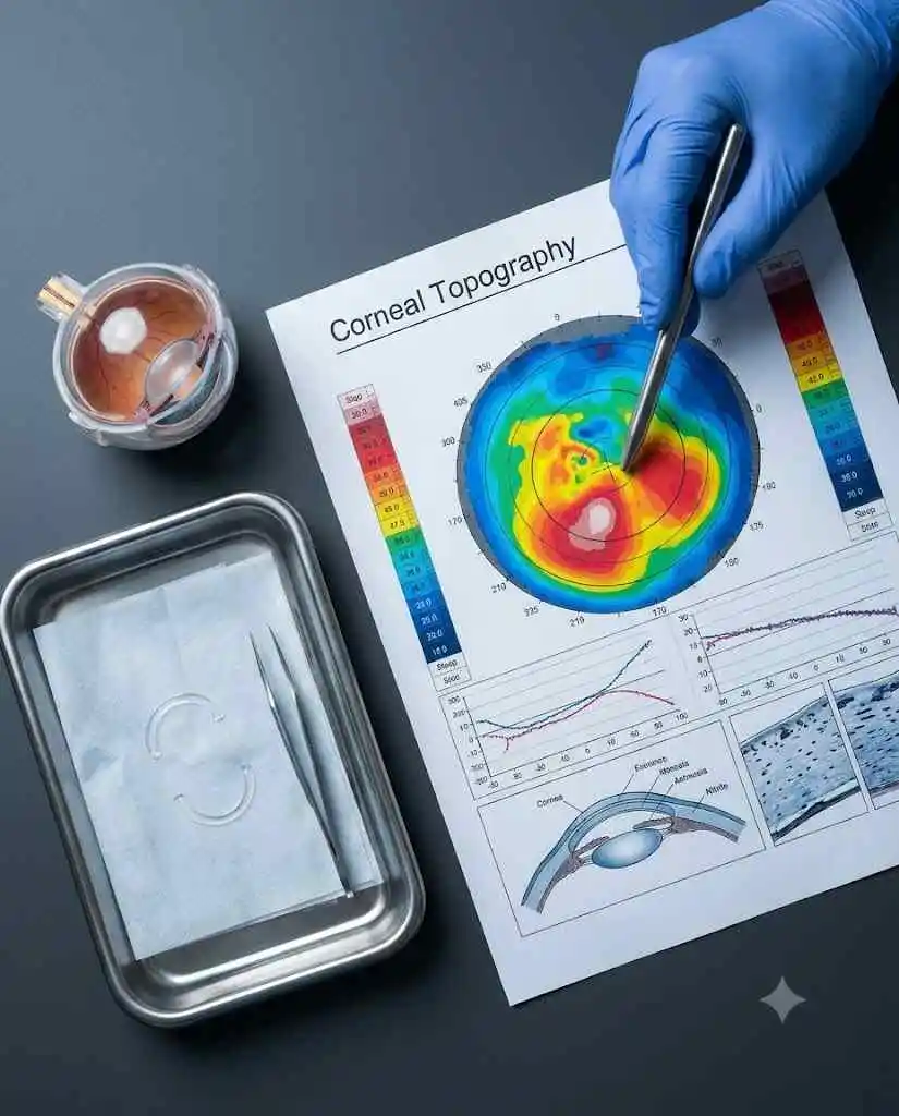
Your surgeon will confirm the diagnosis with corneal topography and a slit-lamp exam. A typical keratoconus pattern shows an asymmetric bow-tie shape plus at least one sign, such as localized thinning, a conical bulge, a Fleischer ring, Vogt striae or surface scarring.
Stability is checked too. In research, stable keratoconus meant no more than 1.00 diopter of change in average curvature over 12 months. Corneal thickness is mapped as well, since it decides which segment sizes are safe.
From these measurements, the surgeon plans how many segments to use, how thick they should be and how long each arc should be. The incision is placed on the steepest meridian of the cornea.
How the Procedure Is Performed, Step by Step
- Numbing drops or local anesthesia keep the eye comfortable.
- A narrow tunnel is created in the corneal stroma, either with a femtosecond laser or a manual dissector.
- The segments are slid into the tunnel through a tiny incision.
- The surgeon checks their position and protects the eye with drops and, often, a shield.
Clinics quote different times for the operation itself, from about 15 minutes per eye to around 30 minutes. The whole visit usually lasts about an hour. The laser method gives more precise control over tunnel depth and size, which is a big reason complication rates have fallen.
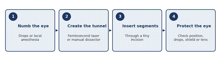
Figure 2. The four main stages of ring implantation (illustration).
Recovery After ICR Eye Surgery: What to Expect
Most people rest with their eyes closed for a few hours afterward, then go home the same day. Recovery is generally much faster than after a corneal transplant.
Protection after surgery differs by clinic. Some use a shield for a few days, while others place a bandage contact lens that stays for up to two weeks. Drops are commonly needed for two to four weeks.
Expect to use prescribed eye drops and to avoid rubbing the eye. Research protocols checked patients at 24 hours and again at one, three and six months.
Vision often improves noticeably by the six-month mark. It can fluctuate early on, and many patients still wear glasses or contact lenses for their sharpest vision.
Everyday Questions: Work, Driving, Lenses and Both Eyes
- Going back to work: Many people resume normal activities within a few days. Some clinics report a return as early as the next day, and one hospital leaflet says patients may go back whenever they feel ready, depending on the job.
- Driving: This depends on how well each eye sees. Wait until your vision is clear and your surgeon gives the go-ahead.
- Swimming and eye rubbing: Skip swimming until your first check-up, and avoid rubbing your eyes.
- Contact lenses: Many people still need lenses afterward, but lenses often fit better once the cornea is more regular. Follow your surgeon’s timing for restarting them.
- One eye or both: Ask whether the eyes should be treated separately. Staggering them can let the untreated eye keep working while the first one heals.
- Age: Early U.S. approval criteria covered patients over 20. Keratoconus tends to stay active during the first three decades of life, so surgeons are extra careful to document stability in younger patients.
What Results Can You Realistically Expect?
The answer depends on whether your cone is stable. In the five-year study of stable keratoconus, gains in vision and refraction seen at six months held steady, and curvature crept back by less than 1.00 diopter.
The picture changes when the disease is active. In a small group of 18 progressing eyes, curvature dropped by 4.40 diopters at six months, yet 3.36 diopters of that benefit had returned by year five. That study was retrospective and small, and some patients may have stabilized naturally, so treat it cautiously.
The takeaway is clear. The rings improve shape and vision, but they do not cure keratoconus. Independent reviewers have also assessed the wider evidence base for ICRS, and that is worth a look if you want more depth.
Combining Rings With Corneal Cross-Linking
Corneal cross-linking uses ultraviolet light to strengthen corneal tissue, while the rings reshape it. Because the two tackle different problems, surgeons sometimes combine them.
A small early study of four eyes shows the idea. The implants gave an average gain of two lines in corrected vision and six lines in uncorrected vision. Cross-linking, performed two to six months later once vision was stable, added roughly one more line of corrected vision.
Two eyes lost some of the ring effect. Swapping a single segment for two thicker ones restored up to three lines. The authors concluded that more research is needed to find the best combination, so ask your surgeon how they sequence the two.
Some centers go a step further. A study at a Korean university followed the rings with laser reshaping of the corneal surface between the segments, plus accelerated cross-linking, usually about 2.7 months later. Such triple approaches remain investigational, so ask whether any apply to your case.
Risks and Possible Complications
ICR eye surgery is considered safe when performed well, largely because femtosecond lasers make tunnels more predictable. Still, no operation is risk-free.
- During surgery: a tunnel that is too shallow or too deep, an off-center segment or an uneven position. Corneal perforation is the most serious event, reported in under 0.6% of laser-assisted cases. Lost suction and minor bleeding on the white of the eye are milder.
- After surgery: infection of the cornea is the most feared problem, seen in under 0.1% of laser-assisted cases. Segment migration or extrusion happens more often with manual tunnels.
- Rare problems: severe glare or halos, a persistent surface defect, inflammation or corneal melting. These can require removal of the segments.
- Harmless findings: white deposits inside the tunnel appear in up to 60% of eyes and do not affect vision or structure.
Reported removal rates vary widely, from 1% to 30%, though large series cluster around 4% to 6%. One long, asymmetric ring design had a 17% removal rate, with five of 30 eyes affected by melting near the incision, so surgeons avoid such designs until safer versions exist.
If a segment must come out, removal is safe, and the cornea tends to drift back toward its original shape. A growing change in astigmatism can be an early warning of late extrusion.
Rings vs. Other Keratoconus Treatments
Here is how the main keratoconus treatment options compare.
| Option | What it does | Main limitation |
| Glasses or soft lenses | Corrects mild blur | Cannot fix an irregular shape |
| Specialty or rigid lenses | Create a smoother optical surface | Comfort and handling problems |
| Corneal cross-linking | Strengthens tissue to slow progression | Aims to stabilize, not improve vision |
| Intracorneal rings | Reshape and regularize the cornea | May not stop progression |
| Corneal transplant | Replaces diseased tissue | Longer recovery and higher cost |
Transplant options include penetrating keratoplasty and lamellar keratoplasty. Compared with a transplant, intracorneal ring segments are less invasive and reversible, recovery is faster and the cost is lower.
Cost and Insurance Coverage in the U.S.
Many U.S. insurers treat intracorneal ring segments for keratoconus as medically necessary. The procedure is billed under CPT code 65785. Some plans also cover ectasia after LASIK or PRK, while others limit coverage to keratoconus.
Prices for corneal ring implants vary by surgeon, facility and segment type. Ask for an itemized estimate and request prior authorization before booking ICR eye surgery.
What the Future Holds: AI and Personalized Planning
Researchers recently tested whether artificial intelligence could pick better intracorneal ring segments. In a pilot of 40 eyes, half were treated using the manufacturer’s nomogram and half using a neural network.
Both groups improved, but the network group did better. Corrected vision moved from 0.20 to 0.15 logMAR, compared with 0.26 to 0.22 in the nomogram group, and higher-order aberrations fell more. The sample was small, so larger trials are needed.
Computer modeling is heading the same way. A 2024 study built an automated simulation and tested 300 variations of 20 ring designs to learn which geometric features best predict results. The aim is to personalize nomograms so fewer patients need revision surgery.
Questions to Ask Before ICR Eye Surgery
- Is my keratoconus stable enough for rings?
- Am I likely to gain vision, given my starting acuity?
- Will you use a femtosecond laser or a manual dissector?
- Which segment type and size suit my cornea, and why?
- Do I need cross-linking as well?
- What would removal involve, and what will it all cost?
Conclusion: Is ICR Eye Surgery Right for You?
ICR eye surgery works best for people with real vision loss, contact lens trouble and a stable cone. It improves corneal shape and often restores useful vision, yet it does not cure keratoconus, and glasses may still be needed.
If this sounds like your situation, book a visit with a corneal specialist who can map your cornea and explain realistic outcomes. This article is for information only and is not a substitute for medical advice.
Frequently Asked Questions
Is ICR eye surgery painful?
Most people feel pressure rather than pain. Anesthetic drops numb the eye, and the operation usually takes about 15 to 30 minutes. Mild scratchiness or light sensitivity afterward is common and usually settles quickly.
How long do corneal ring implants last?
The segments are meant to stay in place. In stable keratoconus, results held for at least five years in research. If the cone keeps progressing, the benefit may fade.
Can intracorneal rings be removed?
Yes. Removal is considered safe, and the cornea gradually moves back toward its earlier shape. Surgeons remove segments for infection, melting, extrusion or troublesome glare.
Will I still need glasses after ICR eye surgery?
Possibly. The rings improve corneal shape, but many patients continue to use glasses or contact lenses for their clearest vision. The difference is that lenses usually become easier to tolerate.
Does insurance cover ICR eye surgery?
Often, yes, for keratoconus, because insurers commonly view it as medically necessary. Coverage for post-LASIK ectasia varies, so confirm your plan’s rules and get prior authorization first.