
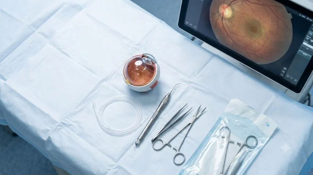
MIVS eye surgery is now the way many retina surgeons carry out a vitrectomy, and it has made the day of the operation and the weeks after it easier on patients. MIVS stands for microincision vitrectomy surgery. The surgeon uses very fine tools through cuts about half a millimetre wide, rather than larger tools and stitches to close the eye wall.
If your doctor has mentioned this operation, you probably want clear answers. Below you will find how MIVS eye surgery works, what it treats, how the instrument sizes compare, the main risks and what healing looks like.
Key Takeaways
- MIVS is a vitrectomy performed with ultra-thin 23-, 25- or 27-gauge instruments.
- The small wounds usually seal on their own, so many patients need no stitches and feel less irritation than after older 20-gauge surgery.
- Surgeons use it for retinal detachment, bleeding from diabetic eye disease, macular holes, macular pucker and other retinal conditions.
- Low eye pressure, infection, retinal tears and cataract can happen, but serious problems are uncommon with experienced surgeons.
- Healing depends on what is being treated. A gas bubble or silicone oil brings head-positioning rules and travel limits.
- The disease being treated shapes the result more than the gauge does, and surgeon experience counts for more than the choice between 23, 25 and 27.
What Is MIVS Eye Surgery?
MIVS eye surgery is a type of vitrectomy. Through three tiny openings, each about 0.6 mm or smaller and usually left unstitched, the surgeon removes the gel inside the eye to treat problems in the retina and vitreous.
Your eye is filled with vitreous, a clear jelly in front of the retina. If that jelly fills with blood, scar tissue or debris, or tugs on the retina, sight gets worse. In a vitrectomy the gel is taken out so the surgeon can deal with the cause and mend the retina beneath.
The microincision method works the same way but with much finer tools. Instrument size is measured in gauge, where a bigger number means a thinner instrument. Most surgeons today use 23-, 25- or 27-gauge systems, so you may hear the term small-gauge vitrectomy.
Three small ports are made in the white of the eye. One supplies fluid that keeps the eye firm, one holds a thin light, and one admits the cutter and other instruments. The surgeon watches through a microscope and a wide viewing lens.
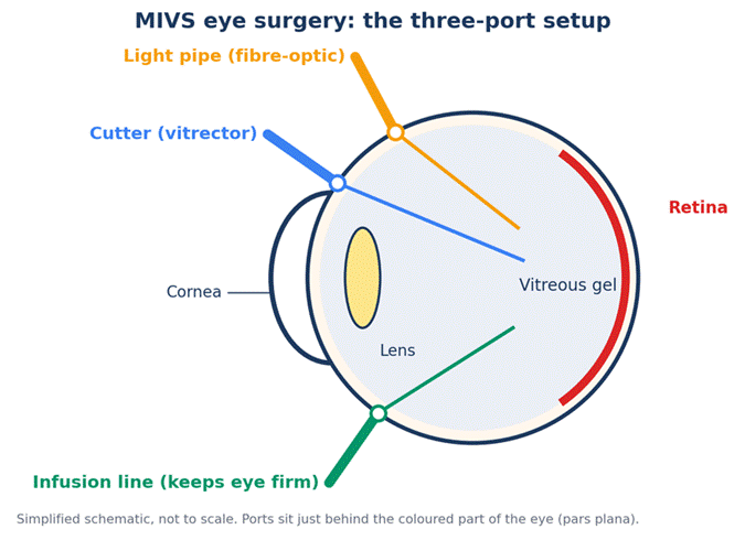
Figure 1. The three ports used during the operation.
How Microincision Vitrectomy Surgery Developed
Modern vitrectomy began in 1971, when Robert Machemer built a closed system for removing the gel. By 1974 the three-port 20-gauge approach, sealed with dissolving stitches, had taken hold and remained the routine method for more than three decades.
Surgeons began moving toward sutureless operations in 2002, using 25-gauge instruments passed through tiny cannulas. Eckardt’s 23-gauge system followed a few years later, and Oshima’s team developed 27-gauge after that. At every stage the tools got thinner and the wounds smaller.
The common gauge sizes compare like this:
| Gauge | Approx. width | What to know |
| 20G | about 0.9 mm | Older standard. Usually needs an opening in the conjunctiva and stitches. |
| 23G | about 0.6 mm | Sturdier tools, widely used and often stitch-free. |
| 25G | about 0.5 mm | First widely used system that passes through the conjunctiva, introduced in 2002. |
| 27G | about 0.4 mm | Smallest in routine use. Bendier, so makers add stiffening sleeves. |
Figure 2. Instrument widths drawn to scale.
Conditions Treated With Small-Gauge Vitrectomy
A vitrectomy is seldom the whole treatment. It opens the way for other work inside the eye. Retina specialists commonly turn to it for:
- Retinal detachment, where the retina peels away from the back wall of the eye
- Diabetic eye disease, including bleeding into the vitreous and scar tissue that pulls on the retina
- Macular hole, a small break in the central retina
- Epiretinal membrane, also called macular pucker, a layer of scar tissue that wrinkles the macula
- Vitreomacular traction, where the gel tugs on the macula
- Eye infections, serious injuries and lens pieces left behind after cataract surgery
In the same operation the surgeon may peel off membranes, seal tears with laser, or fill the eye with air, gas or silicone oil to hold the retina in position.
Warning Signs and How to Prepare
Some retina problems need quick action. A sudden burst of floaters, flashing lights, a dark curtain drifting across your vision or abrupt sight loss calls for an eye exam the same day. Retinal detachment is the best-known example, and the sooner it is repaired the better the retina’s chances.
Once an operation is booked, your team will tell you which medicines to keep taking, including blood thinners, and whether you need to fast. It is normally day surgery, so line up a ride home. When both eyes need surgery, they are done one at a time, with the second eye waiting until the first has healed.
How the Procedure Works, Step by Step
Each case differs, but a typical MIVS eye surgery operation runs in this order:
- Anaesthesia. The eye is cleaned with antiseptic, then most patients get local anaesthetic and light sedation. Long or complicated cases may call for general anaesthesia.
- Port placement. The surgeon nudges the conjunctiva aside and enters at a slant before straightening the instrument. This angled route lets the wound shut like a small valve afterwards.
- Vitreous removal. The cutter takes out the gel in tiny bites, working from the centre outward.
- Repair. Scar tissue is peeled away, tears are sealed with laser and a detached retina is pressed back into place.
- Tamponade. Air, gas or silicone oil is placed in the eye when the retina needs support.
- Closing. The cannulas come out, the wounds are checked for leaks and one stitch is added if a wound looks unsafe.
How long does it take? A simple case such as a macular pucker can take about an hour. Complicated detachments or advanced diabetic disease can take longer, because the time depends mainly on what else must be done inside the eye rather than on the gauge.
Some surgeons take out a cataract during the same session. This saves a second operation and heads off the cataract that vitrectomy tends to bring on sooner, especially after 50.

Benefits of MIVS Eye Surgery Over Older 20-Gauge Surgery
Final outcomes with stitch-free surgery are broadly on par with 20-gauge. The real difference shows up in how comfortable the operation feels, how long it takes and how quickly you heal.
- Gentler first days: The conjunctiva is barely disturbed and no suture ends rub under the eyelid, so many patients report little scratchiness the day after surgery.
- Sharper vision sooner: Tiny wounds distort the front of the eye less, so the astigmatism that follows surgery is usually milder and sight stabilises earlier.
- Shorter overall time: Getting into the eye and sealing it takes far less time, which often compensates for the slower vitreous removal of early small-gauge machines.
- Finer cutting: Newer machines can cut up to 7,500 times a minute, so each bite of gel is smaller and pulls less on the retina.
What the Research Says About Results and Gauge Choice
Outcome depends far more on the disease than on the gauge. A 2020 meta-analysis of 11 studies and 940 eyes compared 27-gauge with 25-gauge surgery. For retinal detachment, about 92 to 93 percent of eyes were repaired with a single operation in both groups, with individual studies falling between roughly 85 and 97 percent.
The 27-gauge operations took about three minutes longer on average, with comparable pressure and vision, and fewer complications overall, mostly in epiretinal membrane cases. The authors noted that the trials were small and that larger studies are needed.
Macular holes show similar results. A review of 211 eyes found that about 95 percent closed after one operation, with air or gas, and all had closed once repeat surgery was counted. Smaller, more recent holes generally do best.
Is one gauge clearly the best? Not according to surgeons. A 2018 Retina Today review noted that 23-, 25- and 27-gauge systems have each earned a place as an accepted standard, while opinions differ on which is superior. What often matters more is how much experience your surgeon has with the system being used.
How much vision returns depends on how damaged the retina was beforehand. In a Bascom Palmer Eye Institute review of 327 eyes treated for macular problems, most patients gained two or more lines of vision with few complications. Older patients with detachments improve too, though usually by less than younger ones.
Is Vitrectomy for Floaters Worth Considering?
Floaters are drifting specks or strands that most people notice at some point, and they are usually harmless. Many fade into the background within a few months, and a reassuring retina check is often enough. Surgery is kept for floaters so dense that they get in the way of reading or driving.
The options are waiting it out, laser or vitrectomy. In a review of 168 eyes treated with 25-gauge tools, vision improved on average from 20/40 to 20/25 and most patients were satisfied. Yet about 7 percent developed a retinal break, and cataract, detachment and infection remain possible. That trade-off explains why, in a 2010 poll at an Academy meeting, about two thirds of ophthalmologists voted against surgery for floaters.
Risks and Complications of Small-Gauge Vitrectomy
No eye operation is risk free. Much of the data below comes from studies in the 2000s, so it reflects early experience and varies between surgeons and eyes.
Low eye pressure (hypotony): A wound may leak a little, especially early on. Some 25-gauge series reported temporary hypotony in up to 20 percent of eyes, usually resolving within days. Surgeons lower the risk with angled wounds, careful cannula removal and, if needed, an air or gas fill.
Infection (endophthalmitis): Early reports tied stitch-free vitrectomy to more infections, and surgeons took that seriously, but later large studies did not confirm the link. Infection after any vitrectomy is rare yet urgent.
Retinal tears and detachment: Breaks can form near the entry sites. Most series report them during surgery in under 5 percent of cases. Detachment afterwards is possible, with rates depending on the condition treated.
Other risks: Slipped cannulas caused choroidal detachment in about 3.5 percent of cases in one review. Pressure spikes, bleeding and silicone oil leaking under the conjunctiva have also been reported. Cataract is the most common long-term effect and is more likely after 50. Occasionally a surgeon adds a stitch, for example if the sclera is thin, the eye has had several operations or a wound leaks.
Recovery and Aftercare After MIVS Eye Surgery
Healing depends on the eye problem and on whether a bubble was used. The National Eye Institute’s patient guide to vitrectomy says the eye can stay red and swollen for several weeks, with some pain and blurry vision early on. Expect a patch for about a day, antibiotic or anti-inflammatory drops, and a pause on driving, hard exercise and heavy lifting. A check-up usually follows the next day to assess pressure and wound sealing, then more visits over the coming weeks.
If a gas bubble is used, your surgeon will explain how to hold your head, often for several days. Silicone oil does not absorb by itself and is usually taken out months later.
Call your surgeon right away for worsening pain, a sudden drop in vision, increasing redness, lid swelling or pus-like discharge. Infection is uncommon, but it tends to appear in the first few days, and the eye feels worse instead of better.
Gas Bubble Timeline: Vision, Driving and Flying
While gas fills the eye, vision is very blurred. As the bubble shrinks you will notice a wavy horizontal line that bobs when you move your head, then a small circle that finally vanishes. Hospital leaflets give rough absorption times of two to four weeks for SF6, four to eight for C2F6 and up to about twelve for C3F8.
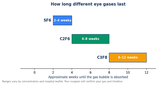
Figure 3. Approximate gas absorption times.
Do not drive while gas or oil is in the eye, and at the very least wait for your first check-up. Flying and high altitudes are also off limits, since gas expands as air pressure drops and can push eye pressure to dangerous levels. Aviation medical guidance suggests roughly two weeks after SF6 and six weeks after C3F8, but you need your surgeon’s clearance. Tell any anaesthetist about the bubble before other procedures, because nitrous oxide must be avoided.
Frequently Asked Questions
What does MIVS stand for in eye surgery?
MIVS stands for microincision vitrectomy surgery, a vitrectomy done through very small incisions, usually with 23-, 25- or 27-gauge instruments.
Is MIVS eye surgery painful?
The operation itself should not hurt, since the eye is numbed and you are sedated. Afterwards you can expect soreness, redness and blurry vision. Many people find it milder than stitched surgery, and your surgeon can prescribe pain relief if needed.
How long is recovery after MIVS eye surgery?
Many people manage light activity within a week or two. The eye can stay red and swollen for several weeks, and full visual recovery can take months, especially with gas or silicone oil. Your own timeline depends on the condition treated.
How long does MIVS eye surgery take?
Straightforward cases often take around an hour, though complex detachments or diabetic cases can take longer. Allow extra time at the surgical centre for preparation and recovery before heading home.
Can MIVS eye surgery cause cataracts?
Yes. Cataract is among the most common effects of any vitrectomy, particularly after 50. If you already have an artificial lens from cataract surgery, vitrectomy does not damage the implant.
Bottom Line
MIVS eye surgery gives retina surgeons a gentler way to treat serious problems inside the eye, with smaller wounds, fewer stitches and often an easier recovery. It is not risk free, and the outcome still depends on the condition and the surgeon’s skill. If a vitrectomy is suggested, ask which gauge is planned, what to expect afterwards and which warning signs to watch for.