
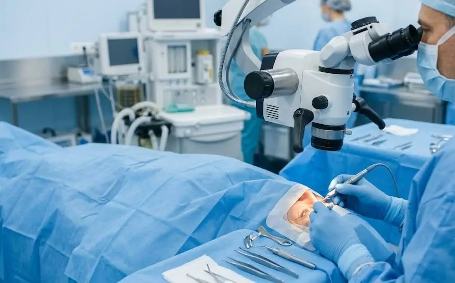
MICS eye surgery is a modern cataract-removal technique that uses an opening of roughly 2 millimetres to remove the cloudy lens and insert a folded artificial lens through that same small entry. If your eye doctor has suggested it, or you have seen it promoted by a clinic, you may be asking how it differs from conventional cataract surgery.
In simple terms, the incision is smaller, which may make the healing process somewhat gentler. This guide explains the more important details: how MICS works, who may be suitable, what research has found, what recovery is like, and the typical cost in India.
Key Takeaways
- MICS means micro incision cataract surgery. A cloudy natural lens is removed through an opening of about 1.8 to 2.2 mm, and stitches are usually unnecessary.
- MICS is a refinement of phacoemulsification, which is the usual cataract procedure, rather than an entirely separate form of treatment. Both approaches can be safe and effective when performed by experienced surgeons.
- A smaller opening can produce slightly less surgically induced astigmatism, although the strongest trial evidence describes the difference as small and the certainty of the evidence as low.
- The type of artificial lens selected has a much greater influence on vision after surgery than the size of the surgical opening.
- Because costs differ considerably according to the city, clinic and lens, request a written quotation showing exactly what you are paying for.
What Is MICS Eye Surgery?
MICS is short for micro incision cataract surgery. It is also commonly described as MICS eye surgery or MICS cataract surgery. The procedure addresses cataract, meaning the natural lens of the eye has become cloudy.
During MICS, ultrasound energy is used to divide the cloudy natural lens into small pieces. This is the phacoemulsification technique. The pieces are then removed by suction and replaced with a clear artificial lens, called an intraocular lens (IOL).
The term “micro” refers to the very small entry wound. It is usually around 1.8 to 2.2 millimetres, approximately the width of a grain of rice. Openings of this size normally seal without help, meaning most patients do not need a stitch.
There are two main forms of MICS eye surgery. Coaxial MICS is the more commonly used approach; the fluid and ultrasound probe pass through one opening measuring about 1.8 to 2.2 mm. With bimanual MICS, or biaxial MICS, these functions use separate openings, each commonly 1.5 mm or less. This permits an even smaller entry, but it requires greater technical skill and the lens may still require a somewhat larger opening. The measurements in this guide mainly describe coaxial MICS.
The abbreviation MICS is also used for procedures outside eye care, including minimally invasive cardiac surgery. In an ophthalmology setting, however, it refers here to the cataract procedure explained in this article.
How Small Is “Micro”? Incision Sizes Compared
A difference of roughly one millimetre may seem insignificant. However, the cornea has a carefully shaped curve, and any incision can alter that shape. In general, a smaller incision causes less disturbance to the cornea.
| Technique | Typical incision | What to know |
| MICS (micro incision) | Approximately 1.8 to 2.2 mm | Requires very thin, foldable lenses and fine instruments. It is generally sutureless. |
| Standard phacoemulsification | Approximately 2.8 to 3.2 mm (some surgeons use smaller openings) | Offers the broadest choice of lenses and is widely available. |
| Femtosecond laser-assisted (FLACS) | Approximately 2.2 to 2.4 mm | A laser carries out some parts of the procedure and the cost is usually higher. |
| Manual small incision (SICS) | Approximately 6 mm or more | The lens is removed in one piece without a phaco machine. It is often used for very hard cataracts. |
The measurements are approximate and can change according to the surgeon, equipment and surgical technique.
How MICS Eye Surgery Is Done, Step by Step
You remain awake during the procedure, which is normally painless. The usual process is as follows.
- Preparation. Eye drops enlarge the pupil, while numbing drops or a local anaesthetic help keep the eye comfortable.
- Making the opening. The surgeon creates a self-closing incision near the edge of the cornea and a very small side opening for another instrument.
- Fragmenting the lens. A narrow ultrasound probe uses low energy to break the cloudy lens into small pieces.
- Removing the fragments. Fluid enters the eye as the broken lens material is suctioned out, helping maintain stable pressure and conditions inside the eye.
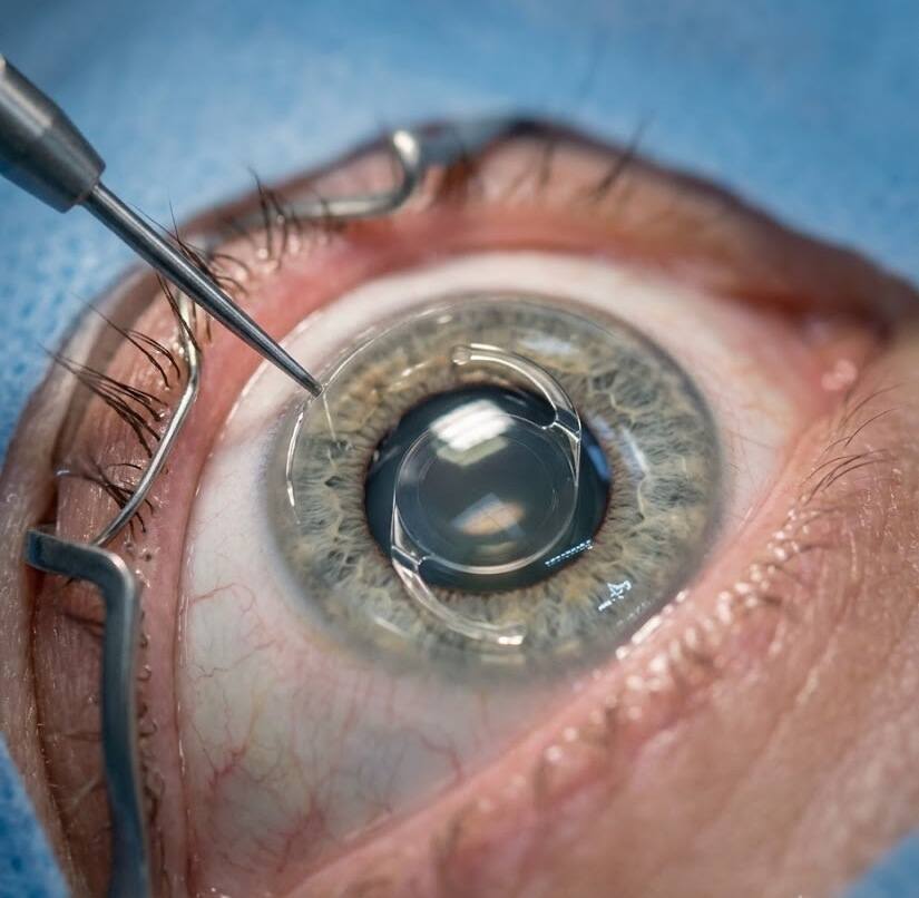
- Inserting the replacement lens. The folded IOL is guided through the same small opening and unfolds inside the thin capsule that previously contained the natural lens.
- Closing the wound. The incision seals on its own. An eye shield is placed for protection, and most patients leave the clinic the same day.
The actual procedure generally lasts well under an hour. Even so, expect to remain at the clinic for a few hours because of the checks carried out before and after surgery.
Who Is a Good Candidate for MICS Eye Surgery?
Age alone does not determine suitability. The more important question is how much the cataract is affecting everyday life. Blurred sight, glare when driving at night, or difficulty reading despite updated glasses are common reasons to discuss surgery with an eye surgeon.
MICS eye surgery may suit you if:
- you would like a toric lens to address astigmatism during the cataract procedure
- you are considering a premium option, such as a multifocal or extended depth-of-focus lens
- you have diabetes or glaucoma and your surgeon considers MICS appropriate for your eye
- you prefer a minimally invasive approach and the clinic has surgeons experienced in MICS
MICS can be less suitable, or may need modification, when the cataract is extremely dense. A surgeon may enlarge the opening slightly to improve safety or select a different surgical approach. This represents a clinical adjustment rather than a failure of the technique.
Before surgery, your evaluation will include eye imaging, measurements of eye length and corneal curvature, plus checks of the retina and eye pressure. These findings—not promotional material—are what should guide the surgical plan.
When Should You Have Cataract Surgery?
Cataracts often develop gradually, so there is usually no reason to hurry. Surgery is commonly recommended once vision begins interfering with everyday activities such as driving, reading, employment or safety. The presence of a cataract by itself does not automatically mean surgery is needed.
Early on, stronger spectacles or improved lighting may provide enough help for some time. Delaying for too long can also create difficulties: a very advanced cataract can make the operation more challenging and, rarely, contribute to other eye problems such as increased eye pressure.
If you are uncertain, ask the surgeon how advanced your cataract has become and what the consequences of waiting could be. That discussion is more useful than following a fixed schedule, whether you choose MICS or another cataract technique.
How to Prepare for MICS Eye Surgery
Preparing for MICS is generally straightforward and can make the day of surgery easier. Your clinic will provide specific instructions, but common preparations include the following:
- Pre-operative tests. Eye scans and measurements of eye length and corneal curvature help determine the required lens power. The clinic may also check blood pressure and blood sugar.
- Tell the surgeon about all medicines you use. Blood thinners are important to mention, as are some prostate medicines, including tamsulosin, which can influence iris behaviour during surgery. Do not stop any medicine without medical advice.
- Contact lenses. You will normally be told to stop using contact lenses before the measurements. Rigid lenses may need to be stopped for around three weeks and soft lenses for around two, although your clinic’s instructions take priority.
- Pre-operative drops. Antibiotic or anti-inflammatory eye drops are commonly begun several days before the operation.
- On the day of surgery. Follow the food and drink instructions from your clinic, avoid makeup and perfume, bring your glasses and medication list, and arrange transportation home.
What Does the Research Actually Show About MICS?
Clinic material may describe MICS as quicker, gentler or more precise. The evidence supports some advantages, but not all of these claims equally well.
What is well established
Phacoemulsification using a foldable IOL remains the standard treatment for age-related cataract and restores useful sight for most patients. This remains true regardless of whether the incision is smaller or larger.
What the comparison studies found
The strongest evidence about incision size comes from a Cochrane systematic review of randomised trials. Eight trials involving nearly 1,000 eyes compared a 2.2 mm opening with a standard incision of about 3.0 mm. At three months, the smaller incision was associated with a small reduction in surgically induced astigmatism, averaging about 0.19 dioptres.
The reviewers judged the certainty of this result to be very low. They also reported that smaller incisions did not show a consistent reduction in astigmatism in every comparison.
Another meta-analysis of randomised trials published in 2017 reported a more favourable result for astigmatism. It nevertheless found no difference between the approaches for safety outcomes or the length of the operation.
For perspective, 0.19 dioptres is less than the 0.25-dioptre increments commonly used in spectacle prescriptions. Most people would probably not notice a difference that small.
What is not established
- A safety advantage has not been established. The meta-analysis described above found no safety difference between micro-incision surgery and standard phaco.
- A sharper final visual result has not been established either. The selected IOL, the condition of the eye and the surgeon’s skill have much greater influence.
- It has also not been proven that every patient heals or becomes comfortable faster. Faster comfort is plausible and frequently reported, but the evidence is less certain than the finding concerning astigmatism.
Put simply, the smaller opening is a useful refinement that offers a modest potential benefit. It does not represent a completely different level of cataract surgery.
Benefits and Limitations of MICS Eye Surgery at a Glance
| Potential benefits | Limitations to keep in mind |
| Very small, usually self-sealing wound with few or no stitches | Some centres do not have the required instruments or training |
| May produce slightly less induced astigmatism | The measured benefit compared with standard phaco is small |
| Compatible with toric and many premium lenses | Ultra-thin foldable lenses are required, which can limit lens choices |
| Can be used for eyes with diabetes or glaucoma when the surgeon considers it appropriate | It may cost more, and dense cataracts may require a larger incision |
Recovery After MICS Eye Surgery: What to Expect
Most patients are reasonably comfortable within the first day or two. Vision can continue to stabilise over several weeks. A common recovery pattern is:
- First 24 hours: sight may be hazy and the eye can feel gritty. Use the protective shield as instructed and avoid rubbing the eye.
- Days 2 to 7: clarity generally improves. With the surgeon’s approval, many people can return to light desk-based work after two or three days.
- Weeks 2 to 4: prescribed drops often continue for around four weeks. Until your surgeon clears you, avoid swimming, dusty environments and strenuous lifting.
- Around one month: healing is largely advanced, and a final spectacle prescription can be measured if you still need glasses.
Follow-up is commonly scheduled within the first day or two, again at roughly one week, and then around one month.
Use antibiotic and anti-inflammatory drops exactly as directed. Skipping doses can increase the risk of preventable complications.
Contact your surgeon promptly if you develop a sudden loss of vision, worsening pain, increasing redness, new flashes, or the appearance of a dark curtain over your sight.
Both Eyes and Daily Life After MICS Eye Surgery
Many surgeons treat the eyes separately, with several days to a few weeks between operations. This allows the first eye to heal and gives the surgeon an opportunity to assess the result before operating on the second. Some centres perform both eyes on the same day, although many surgeons prefer a staged approach.
For normal activities, follow your surgeon’s individual instructions. In general, the following patterns are common:
- Driving: wait for clearance at a follow-up appointment and make sure your vision is comfortable and safe.
- Flying: short air travel is often possible after roughly one week when the eye pressure is normal at review.
- Washing your face and hair: during the first few days, prevent soap and water from entering the eye. Confirm the exact timing with your clinic.
- Eye makeup: it is generally best avoided for the first one to two weeks.
- Screens and reading: these are usually acceptable once vision feels comfortable. Take regular breaks because dryness can occur.
Risks and Side Effects of MICS Cataract Surgery
Serious problems are uncommon, although every operation carries some risk. The complications associated with MICS cataract surgery are broadly similar to those seen with standard phaco:
- Infection inside the eye. This is rare but serious and requires urgent medical treatment.
- Inflammation, swelling or a temporary increase in eye pressure.
- Posterior capsule opacification (PCO). A cloudy layer can develop behind the artificial lens months or years later. A brief laser treatment can clear it.
- Dry eye and sensitivity to light. These effects are usually temporary.
- Retinal detachment or an artificial lens becoming displaced from the centre. Both are uncommon.
Because MICS uses finer instruments and calls for precise technique, the surgeon’s experience with the procedure is relevant. Asking about that experience is reasonable.
Your Lens Choice Matters as Much as the Incision
As with other cataract procedures, the IOL used during MICS has a major effect on the vision you have afterwards. Common choices include:
- Monofocal: provides clear focus at one main distance, usually far away. Reading glasses are still needed by most people.
- Toric: also corrects astigmatism, which may reduce the need for distance glasses.
- Multifocal or extended depth of focus (EDOF): intended to reduce reliance on spectacles, although halos or night-time glare can occur in some patients.
A premium IOL is not suitable for every eye. Your surgeon should discuss its advantages as well as its limitations and trade-offs.
MICS Eye Surgery Cost in India
There is no single standard price. Estimates published by clinics in India place MICS at about ₹45,000 to ₹90,000 per eye, with the final amount influenced by the lens and city. Some hospitals do not publish prices and instead provide quotations for the specific location.
These factors move the price most:
- the IOL selected (monofocal generally at the lower end, with toric or multifocal options costing more)
- single-use micro tips and cartridges, which increase equipment expenses
- the surgeon’s experience and the hospital’s location and available facilities
- the scans and other tests performed before surgery
Ask the clinic to clarify whether the quotation covers testing, the IOL, medicines and follow-up appointments. Also check with your insurer about cataract coverage and whether premium lenses are excluded.
5 Questions to Ask Your Surgeon Before MICS Eye Surgery
- How many MICS procedures have you performed, and is my cataract appropriate for this approach?
- Which IOL would you recommend for my eye, and what are the advantages and disadvantages?
- Will I need glasses afterwards, and for which activities?
- What services and items are covered by the quoted price?
- If the incision has to be enlarged or the surgical plan changes, what would happen?
Frequently Asked Questions
What is MICS eye surgery and what is its full form?
MICS eye surgery is cataract removal performed through a very small opening, generally 1.8 to 2.2 mm wide. MICS stands for micro incision cataract surgery. The cloudy lens is broken into pieces and removed, followed by insertion of a foldable artificial lens through the same opening.
Is MICS eye surgery better than standard phaco?
Not necessarily. Both techniques can be safe and effective. Research indicates that the smaller incision may reduce astigmatism by a small amount, but the difference is modest and the evidence has low certainty. Suitability depends on the individual eye, cataract density, planned IOL and the surgeon’s experience.
Is MICS eye surgery painful?
Usually not. Numbing eye drops or a local anaesthetic allow you to remain awake while keeping the eye comfortable. For a day or two afterwards, some people experience grittiness or mild irritation, which generally settles quickly.
How long is the recovery after MICS eye surgery?
Many patients feel more comfortable within one to three days, while vision commonly continues to settle over one to four weeks. Eye drops often remain necessary for about a month. Your surgeon should advise when swimming, exercise and heavy lifting are safe.
How much does MICS eye surgery cost in India?
Published figures are approximately ₹45,000 to ₹90,000 per eye, depending on the city, hospital and IOL. Choosing a premium lens can increase the cost. Request a written quotation that clearly states what is included.
Bottom Line
MICS eye surgery is an established refinement of modern cataract surgery. Its very small incision may reduce astigmatism slightly and can support a comfortable recovery, but current research indicates that the overall advantage is modest rather than dramatic.
The key priorities are a thorough assessment, an IOL that matches your visual needs and a surgeon whose experience you are comfortable with. When cataract symptoms begin interfering with daily activities, arrange an eye examination and discuss whether MICS or standard phaco is appropriate for your eye.
This article is general information and does not replace a personal eye examination.